. 2021 Mar 1;20(1):43.
doi: 10.1186/s12943-021-01332-8.

CircWAC induces chemotherapeutic resistance in triple-negative breast cancer by targeting miR-142, upregulating WWP1 and activating the PI3K/AKT pathway

Affiliations

CircWAC induces chemotherapeutic resistance in triple-negative breast cancer by targeting miR-142, upregulating WWP1 and activating the PI3K/AKT pathway

Lei Wang et al. Mol Cancer. .

Abstract

Background: Chemotherapeutic resistance is the main cause of clinical treatment failure and poor prognosis in triple-negative breast cancer (TNBC). There is no research on chemotherapeutic resistance in TNBC from the perspective of circular RNAs (circRNAs).

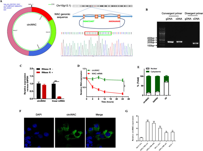
Methods: TNBC-related circRNAs were identified based on the GSE101124 dataset. Quantitative reverse transcription PCR was used to detect the expression level of circWAC in TNBC cells and tissues. Then, in vitro and in vivo functional experiments were performed to evaluate the effects of circWAC in TNBC.

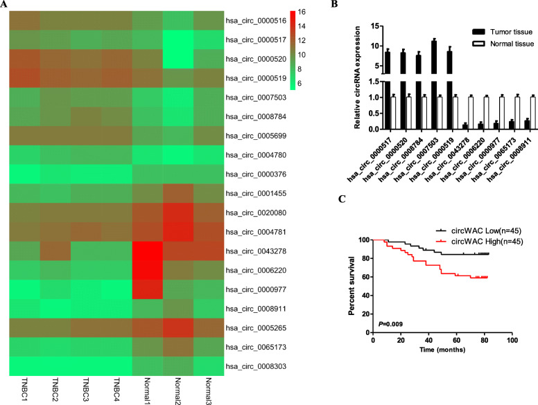
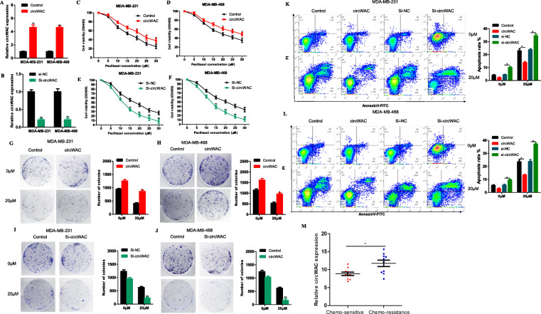
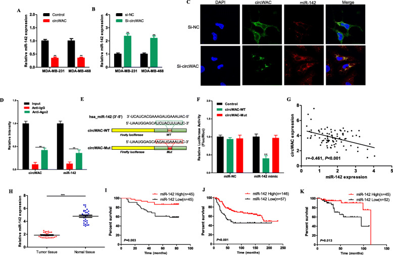
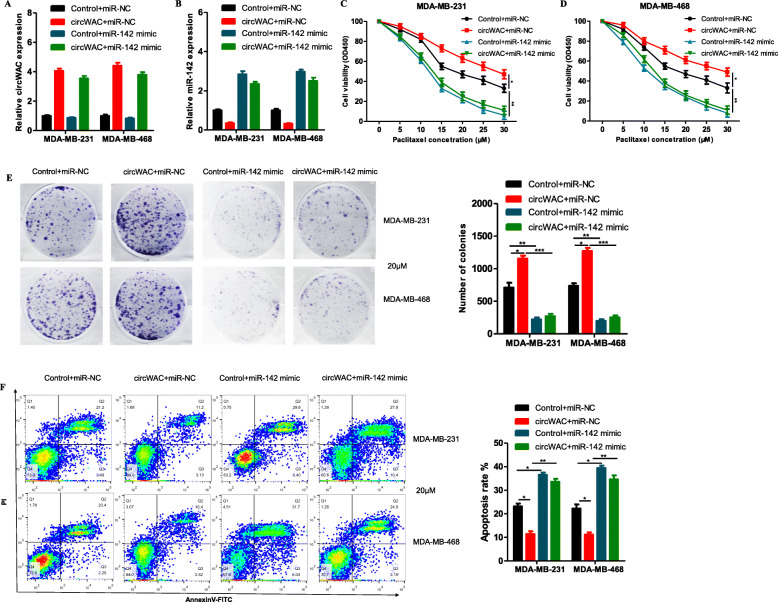
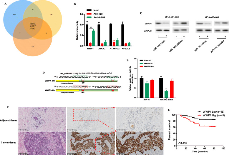
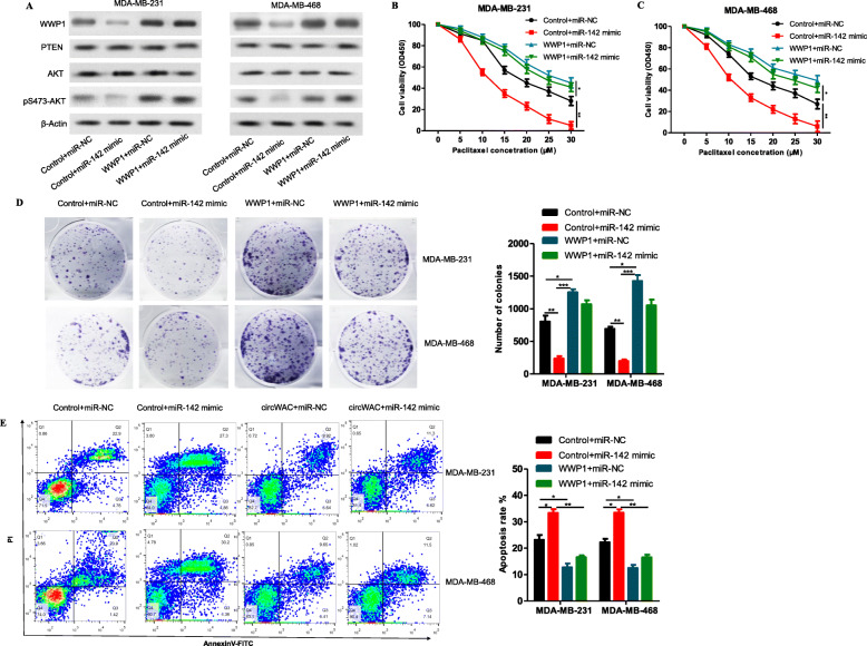
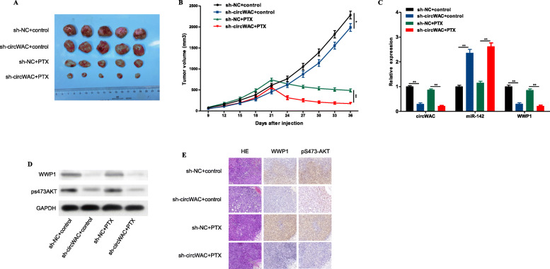
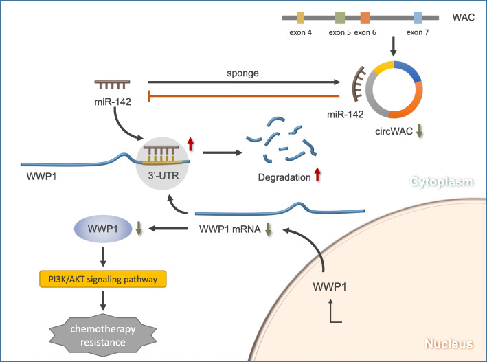
Results: CircWAC was highly expressed in TNBC and was associated with worse TNBC patient prognosis. Subsequently, it was verified that downregulation of circWAC can increase the sensitivity of TNBC cells to paclitaxel (PTX) in vitro and in vivo. The expression of miR-142 was negatively correlated with circWAC in TNBC. The interaction between circWAC and miR-142 in TNBC cells was confirmed by RNA immunoprecipitation assays, luciferase reporter assays, pulldown assays, and fluorescence in situ hybridization. Mechanistically, circWAC acted as a miR-142 sponge to relieve the repressive effect of miR-142 on its target WWP1. In addition, the overall survival of TNBC patients with high expression of miR-142 was significantly better than that of patients with low expression of miR-142, and these results were verified in public databases. MiR-142 regulated the expression of WWP1 and the activity of the PI3K/AKT pathway. It was confirmed that WWP1 is highly expressed in TNBC and that the prognosis of patients with high WWP1 expression is poor.

Conclusions: CircWAC/miR-142/WWP1 form a competing endogenous RNA (ceRNA) network to regulate PI3K/AKT signaling activity in TNBC cells and affect the chemosensitivity of cells.

Keywords: PI3K/AKT; TNBC; WWP1; circWAC; miR-142.


Conflict of interest statement

The authors declare that they have no competing interests.

Figures

Fig. 1
Fig. 1
The expression and prognostic significance of circWAC in TNBC. a The heat map shows the 19 most upregulated and downregulated circRNAs in TNBC tissues compared with normal breast tissues in the GSE101124 dataset. b The top five upregulated circRNAs and top five downregulated circRNAs in TNBC tissues were verified by qRT-PCR. c Kaplan-Meier survival curves indicating the correlation between circWAC and overall survival in TNBC. *P < 0.05, **P < 0.01, ***P < 0.001
Fig. 2
Fig. 2
Characterization of circWAC as a circRNA in TNBC. a Information about the exonic structure of circWAC is illustrated as indicated. The specific primers used for validation of circWAC by Sanger sequencing. b qRT-PCR products generated with divergent primers showing circularization of circWAC. cDNA, complementary DNA. gDNA, genomic DNA. c qRT–PCR analysis of the expression of circWAC and WAC mRNA in MDA-MB-231 cells after treatment with RNase R. d qRT–PCR analysis of the expression of circWAC and WAC mRNA in MDA-MB-231 cells after treatment with actinomycin D at the indicated time points. e Nucleocytoplasmic fractionation experiment showing that circWAC was mainly distributed in the cytoplasm. f FISH analysis of circWAC. Nuclei were stained with DAPI. g The expression level of circWAC in a normal breast cell line (MCF-10A) and breast cancer cell lines was determined by qRT-PCR. The data are presented as the means±S.D. of at least three independent experiments. *P < 0.05, **P < 0.01, ***P < 0.001
Fig. 3
Fig. 3
CircWAC decrease chemosensitivity to PTX in TNBC. a, b The efficiency of circWAC overexpression and silencing in MDA-MB-231 and MDA-MB-468 cells was verified by qRT-PCR. c-f Cells were treated with PTX (concentrations of 0, 5, 10, 15, 20, 25 and 30 μmol/L) for 24 h, and cell viability was evaluated by a CCK-8 assay. g-j Cells were treated with paclitaxel (0 and 20 μM) for 24 h, and cell viability was evaluated by a colony formation assay. k, l Cells were treated with paclitaxel (0 and 20 μM) for 24 h, and the apoptosis rate was analyzed via flow cytometry. The data are presented as the means±S.D. of at least three independent experiments. m The expression of circWAC was significantly increased in the chemotherapy resistance patients. *P < 0.05, **P < 0.01, ***P < 0.001
Fig. 4
Fig. 4
CircWAC acts as a sponge of miR-142 in TNBC. a The expression level of miR-142 in MDA-MB-231 and MDA-MB-468 cells transfected with circWAC or control. b The expression level of miR-142 in MDA-MB-231 and MDA-MB-468 cells transfected with si-circWAC or si-NC. c FISH showed that circWAC and miR-142 were colocalized in the cytoplasm of MDA-MB-231 cells. d The coprecipitated miR-142 was subjected to qRT-PCR to assess circWAC binding by RIP experiments in MDA-MB-231 cells. e MiR-142 and wild-type/mutant circWAC binding site sequences. f Luciferase activity in MDA-MB-231 cells cotransfected with a luciferase reporter vector containing the wild-type or mutant circWAC sequence and the miR-142 mimic or control. g The expression of miR-142 was negatively correlated with circWAC in 90 TNBC patients. h The expression level of miR-142 in TNBC tissues and adjacent nontumor tissues was measured by qRT-PCR. Kaplan-Meier survival analysis of patients with TNBC stratified by the level of miR-142 were used to evaluate OS based on our dataset (N = 90) (I), a METABRIC dataset (N = 203) (J) and a TCGA dataset (N = 97) (K)
Fig. 5
Fig. 5
CircWAC regulates PTX resistance by targeting miR-142. a, b The expression of circWAC and miR-142 in MDA-MB-231 and MDA-MB-468 cells transfected with control + miR-NC, circWAC + miR-NC, control+miR-142 mimic, or circWAC+miR-142 mimic was verified by qRT-PCR. c, d MDA-MB-231 and MDA-MB-468 cells transfected with control + miR-NC, circWAC + miR-NC, control+miR-142 mimic, or circWAC+miR-142 mimic were treated with PTX (concentrations of 0, 5, 10, 15, 20, 25 and 30 μmol/L) for 24 h, and cell viability was evaluated by a CCK-8 assay. e MDA-MB-231 and MDA-MB-468 cells transfected with control + miR-NC, circWAC + miR-NC, control+miR-142 mimic, or circWAC+miR-142 mimic were treated with 20 μM paclitaxel for 24 h, and cell viability was evaluated by a colony formation assay. f MDA-MB-231 and MDA-MB-468 cells transfected with control + miR-NC, circWAC + miR-NC, control+miR-142 mimic, or circWAC+miR-142 mimic were treated with 20 μM paclitaxel for 24 h, and the apoptosis rates were analyzed via flow cytometry. The data are presented as the means±S.D. of at least three independent experiments. *P < 0.05, **P < 0.01, ***P < 0.001
Fig. 6
Fig. 6
WWP1 is the direct target gene of miR-142. a TargetScan, miRDB and miRNAMAP were used to predict the potential target genes of miR-142. b RIP experiments were performed in MDA-MB-231 cells, and the coprecipitated miRNA was subjected to qRT-PCR to evaluate mRNA binding. c The protein expression of WWP1 in MDA-MB-231 and MDA-MB-468 cells transfected with the miR-142 mimic or miR-142 inhibitor was measured by Western blotting. d MiR-142 and wild-type/mutant WWP1 binding site sequences. e Luciferase activity in MDA-MB-231 cells cotransfected with a luciferase reporter vector containing the wild-type or mutant WWP1 sequence and the miR-142 mimic or control. f H&E staining was performed to evaluate tissue morphology, and IHC was performed to evaluate WWP1 expression in TNBC tissue and adjacent tissue. g Kaplan-Meier survival curves indicating the correlation between WWP1 expression and overall survival in TNBC. *P < 0.05, **P < 0.01, ***P < 0.001
Fig. 7
Fig. 7
MiR-142 increases PTX sensitivity and inhibits the PI3K/AKT signaling pathway by targeting WWP1. a The protein levels of WWP1, PTEN, AKT and pS473-AKT in MDA-MB-231 and MDA-MB-468 cells transfected with control + miR-NC, control + miR-142 mimic, WWP1 + miR-NC, or WWP1 + miR-142 mimic were measured by Western blotting. b, c MDA-MB-231 and MDA-MB-468 cells transfected with control + miR-NC, control + miR-142 mimic, WWP1 + miR-NC, or WWP1 + miR-142 mimic were treated with PTX (concentrations of 0, 5, 10, 15, 20, 25 and 30 μmol/L) for 24 h, and cell viability was evaluated by a CCK-8 assay. d MDA-MB-231 and MDA-MB-468 cells transfected with control + miR-NC, control + miR-142 mimic, WWP1 + miR-NC, or WWP1 + miR-142 mimic were treated with 20 μM paclitaxel for 24 h, and cell viability was evaluated by a colony formation assay. e MDA-MB-231 and MDA-MB-468 cells transfected with control + miR-NC, control + miR-142 mimic, WWP1 + miR-NC, or WWP1 + miR-142 mimic were treated with 20 μM paclitaxel for 24 h, and the apoptosis rates were analyzed via flow cytometry. The data are presented as the means±S.D. of at least three independent experiments. *P < 0.05, **P < 0.01, ***P < 0.001
Fig. 8
Fig. 8
A summary map to illustrate the interaction relation among these molecules and signal pathways to drug resistance in TNBC
Fig. 9
Fig. 9
Inhibition of circWAC can increase chemosensitivity to PTX in vivo. a Image of representative resected tumors from four groups of xenografted nude mice on the 36th day. b Tumor volumes were monitored during the 36-d time course. c The expression levels of circWAC, miR-142 and WWP1 in tumor tissues were determined by qRT-PCR. d The levels of WWP1 and pS473-AKT in tumor tissues were detected by Western blotting. e H&E staining was performed to evaluate tissue morphology, and IHC was performed to visualize WWP1 and pS473-AKT. *P < 0.05, **P < 0.01, ***P < 0.001

Similar articles

Cited by

References

    1. Bray F, Ferlay J, Soerjomataram I, Siegel RL, Torre LA, Jemal A. Global cancer statistics 2018: GLOBOCAN estimates of incidence and mortality worldwide for 36 cancers in 185 countries. CA Cancer J Clin. 2018;68:394–424. doi: 10.3322/caac.21492. - DOI - PubMed
    1. Chen W, Sun K, Zheng R, Zeng H, Zhang S, Xia C, Yang Z, Li H, Zou X, He J. Cancer incidence and mortality in China, 2014. Chin J Cancer Res. 2018;30:1–12. doi: 10.21147/j.issn.1000-9604.2018.01.01. - DOI - PMC - PubMed
    1. Fan L, Strasser-Weippl K, Li JJ, St Louis J, Finkelstein DM, Yu KD, Chen WQ, Shao ZM, Goss PE. Breast cancer in China. Lancet Oncol. 2014;15:e279–e289. doi: 10.1016/S1470-2045(13)70567-9. - DOI - PubMed
    1. Chen W, Zheng R, Baade PD, Zhang S, Zeng H, Bray F, Jemal A, Yu XQ, He J. Cancer statistics in China, 2015. CA Cancer J Clin. 2016;66:115–132. doi: 10.3322/caac.21338. - DOI - PubMed
    1. Yoon HJ, Jun CD, Kim JM, Rim GN, Kim HM, Chung HT. Phorbol ester synergistically increases interferon-gamma-induced nitric oxide synthesis in murine microglial cells. Neuroimmunomodulation. 1994;1:377–382. doi: 10.1159/000097191. - DOI - PubMed

Publication types

MeSH terms

LinkOut - more resources