. 2021 Mar 1;40(1):88.
doi: 10.1186/s13046-021-01879-w.

Overexpressed WDR3 induces the activation of Hippo pathway by interacting with GATA4 in pancreatic cancer

Affiliations

Overexpressed WDR3 induces the activation of Hippo pathway by interacting with GATA4 in pancreatic cancer

Wenjie Su et al. J Exp Clin Cancer Res. .

Abstract

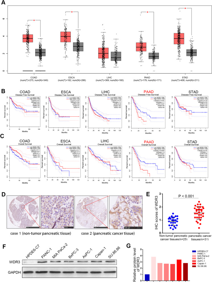
Background: WD repeat domain 3 (WDR3) is involved in a variety of cellular processes including gene regulation, cell cycle progression, signal transduction and apoptosis. However, the biological role of WDR3 in pancreatic cancer and the associated mechanism remains unclear. We seek to explore the immune-independent functions and relevant mechanism for WDR3 in pancreatic cancer.

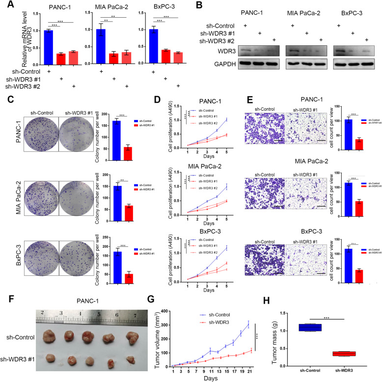
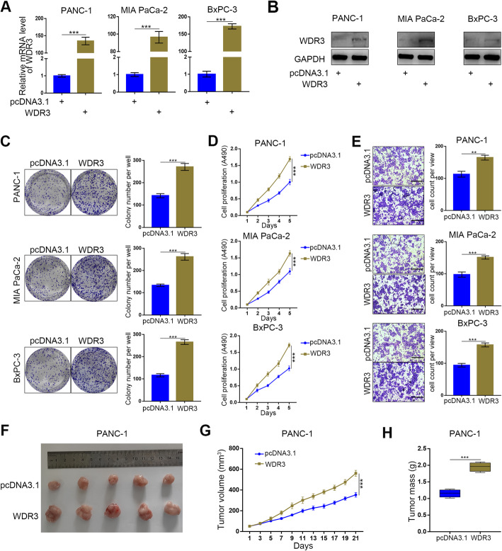
Methods: The GEPIA web tool was searched, and IHC assays were conducted to determine the mRNA and protein expression levels of WDR3 in pancreatic cancer patients. MTS, colony formation, and transwell assays were conducted to determine the biological role of WDR3 in human cancer. Western blot analysis, RT-qPCR, and immunohistochemistry were used to detect the expression of specific genes. An immunoprecipitation assay was used to explore protein-protein interactions.

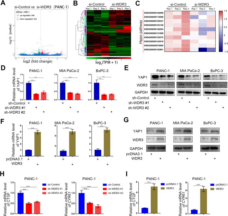
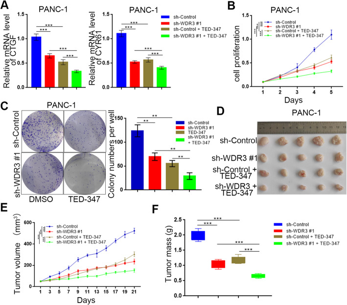
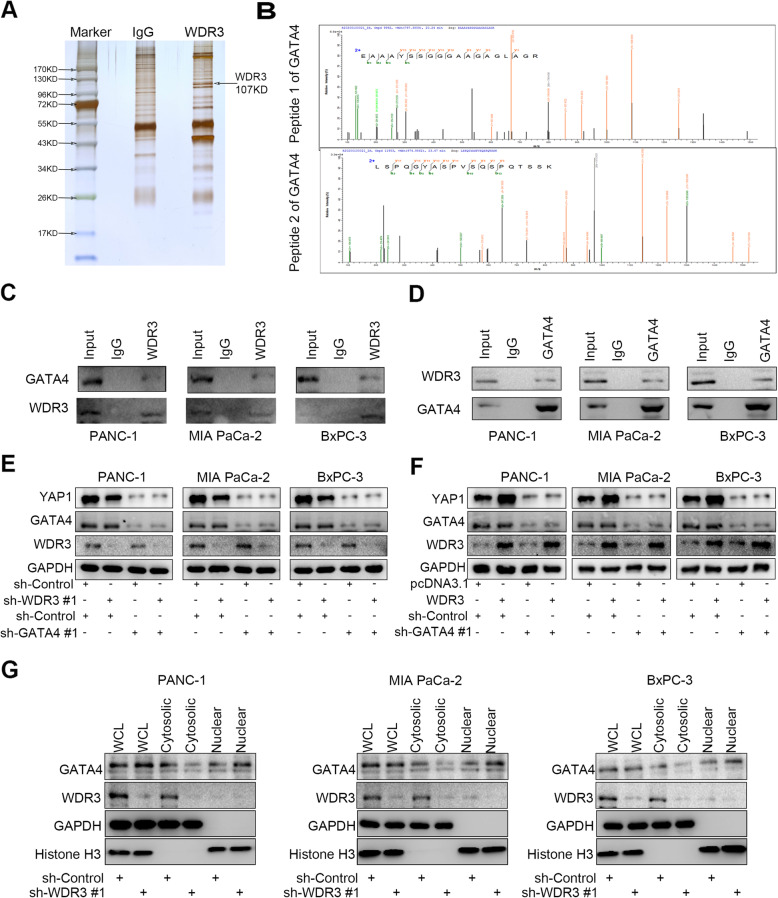
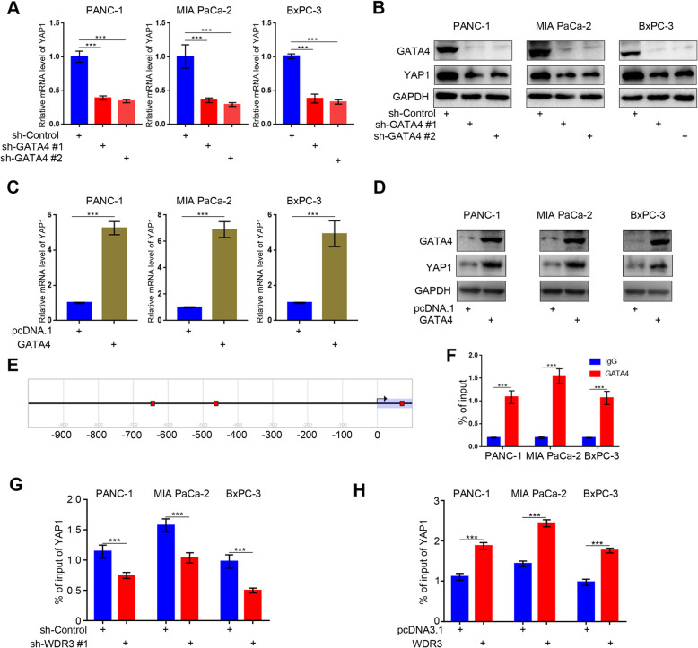
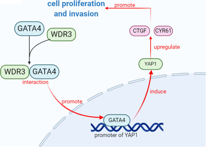
Results: Our study proved that overexpressed WDR3 was correlated with poor survival in pancreatic cancer and that WDR3 silencing significantly inhibited the proliferation, invasion, and tumor growth of pancreatic cancer. Furthermore, WDR3 activated the Hippo signaling pathway by inducing yes association protein 1 (YAP1) expression, and the combination of WDR3 silencing and administration of the YAP1 inhibitor TED-347 had a synergistic inhibitory effect on the progression of pancreatic cancer. Finally, the upregulation of YAP1 expression induced by WDR3 was dependent on an interaction with GATA binding protein 4 (GATA4), the transcription factor of YAP1, which interaction induced the nuclear translocation of GATA4 in pancreatic cancer cells.

Conclusions: We identified a novel mechanism by which WDR3 plays a critical role in promoting pancreatic cancer progression by activating the Hippo signaling pathway through the interaction with GATA4. Therefore, WDR3 is potentially a therapeutic target for pancreatic cancer treatment.

Keywords: GATA4; Hippo signaling pathway; Pancreatic Cancer; WDR3; YAP1.


Conflict of interest statement

The authors have declared that no competing interest exists.

Figures

Fig. 1
Fig. 1
Overexpression of WDR3 is correlated with an unfavorable prognosis in pancreatic cancer. a The GEPIA database was searched for WDR3 mRNA expression in several malignant cancers. Colon adenocarcinoma (COAD), esophageal carcinoma (ESCA), liver hepatocellular carcinoma (LIHC), pancreatic adenocarcinoma (PAAD), and stomach adenocarcinoma (STAD). *, P < 0.05. b-c The GEPIA web tool was searched for disease-free survival (b) and overall survival (c) data for several malignant cancer patients with a high or low WDR3 expression level. P values are shown in the Fig. d. IHC images of WDR3 staining in TMA tissue sections are shown. The scale bars are shown in the Fig. e. Dot plots show the IHC scores of WDR3 expression for TMA tissue sections (normal pancreatic specimens: n = 25, PAAD TMA specimens: n = 31, P < 0.001). Statistical analyses were performed with D’Agostino & Pearson omnibus normality test. f and g Western blot analysis evaluated the expression of WDR3 in normal pancreatic ductal epithelial cells and pancreatic cancer cells (f). The protein expression levels of WDR3 were quantified with ImageJ software (g). GAPDH served as an internal reference
Fig. 2
Fig. 2
Silencing WDR3 suppresses the aggressive behavior of pancreatic cancer cells in vitro and pancreatic tumor growth in vivo. a and b RT-PCR (a) and western blot analyses (b) of WDR3 expression in PANC-1, MIA PaCa-2, and BxPC-3 cells infected with sh-Control or sh-WDR3s. GAPDH served as an internal reference. Data are presented as the mean ± SD of three independent experiments. Each sh-WDR3 group was compared with sh-Control group. Statistical analyses were performed with one-way ANOVA followed by Tukey’s multiple comparison’s tests. **, P < 0.01; ***, P < 0.001. C-E. PANC-1, MIA PaCa-2, and BxPC-3 cells were infected with sh-Control or sh-WDR3 #1. The cells were harvested for colony formation (c), MTS (d), and Transwell invasion assays (e) after 48 h of culture. Each bar represents the mean ± SD of three independent experiments. **, P < 0.01; ***, P < 0.001. F-H. PANC-1 cells infected with sh-Control or sh-WDR3 #1 were subcutaneously injected into nude mice. The tumors were harvested and photographed (f) on day 21. Data for tumor volume (g) and tumor mass (h) are shown as the mean ± SD (n = 5). Each sh-WDR3 group was compared with sh-Control group. Statistical analyses were performed with one-way ANOVA followed by Tukey’s multiple comparison’s tests. ***, P < 0.001
Fig. 3
Fig. 3
Overexpressed WDR3 promoted the aggressive behavior of pancreatic cancer cells in vitro and pancreatic tumor growth in vivo. a and b RT-PCR (a) and western blot analyses (b) of WDR3 expression in PANC-1, MIA PaCa-2, and BxPC-3 cells infected with pcDNA3.1 or WDR3 plasmid. GAPDH served as an internal reference. Data are presented as the mean ± SD of three independent experiments. WDR3 group was compared with pcDNA3.1 group. Statistical analyses were performed with one-way ANOVA followed by Tukey’s multiple comparison’s tests. ***, P < 0.001. C-E. PANC-1, MIA PaCa-2, and BxPC-3 cells were infected with pcDNA3.1 or WDR3 plasmid. The cells were harvested for colony formation (c), MTS (d), and Transwell invasion assays (e) after 48 h of culture. Each bar represents the mean ± SD of three independent experiments. **, P < 0.01; ***, P < 0.001. F-H. PANC-1 cells infected with pcDNA3.1 or WDR3 plasmid were subcutaneously injected into nude mice. The tumors were harvested and photographed (f) on day 21. Data for tumor volume (g) and tumor mass (h) are shown as the mean ± SD (n = 5). WDR3 group was compared with pcDNA3.1 group. Statistical analyses were performed with one-way ANOVA followed by Tukey’s multiple comparison’s tests. ***, P < 0.001
Fig. 4
Fig. 4
WDR3 transcriptionally increases YAP1 expression in pancreatic cancer cells. a and b. Volcano plot (a) and heatmap (b) showing the differentially expressed genes in PANC-1 cells infected with si-Control or si-WDR3. The blue points represent the downregulated genes (n = 248), while the red points represent the upregulated genes (n = 242). c Heatmap showing a subset of WDR3 knockdown-regulated genes with a p-value < 0.1 participating in the Hippo signaling pathway in PANC-1 cells. si-WDR3 group was compared with si-Control group. Statistical analyses were performed with one-way ANOVA followed by Tukey’s multiple comparison’s tests. d and e RT-PCR analysis (d) and western blot analysis (e) to detect the mRNA and protein expression levels of YAP1 in pancreatic cancer cells infected with sh-Control or sh-WDR3s. GAPDH served as an internal reference. Data are shown as the mean ± SD (n = 3). Statistical analyses were performed with one-way ANOVA followed by Tukey’s multiple comparison’s tests. **, P < 0.01; ***, P < 0.001. F-G. RT-PCR analysis (f) and western blot analysis (g) to detect the mRNA and protein expression levels of YAP1 in pancreatic cancer cells infected with pcDNA3.1 or WDR3. GAPDH served as an internal reference. Data are shown as the mean ± SD (n = 3). Statistical analyses were performed with one-way ANOVA followed by Tukey’s multiple comparison’s tests. ***, P < 0.001. h RT-PCR analysis to detect the mRNA expression levels of CTGF and CYR61 in pancreatic cancer cells infected with sh-Control or sh-WDR3s. GAPDH served as an internal reference. Data are shown as the mean ± SD (n = 3). Statistical analyses were performed with one-way ANOVA followed by Tukey’s multiple comparison’s tests. ***, P < 0.001. i RT-PCR analysis to detect the mRNA expression levels of CTGF and CYR61 in pancreatic cancer cells infected with pcDNA3.1 or WDR3. GAPDH served as an internal reference. Data are shown as the mean ± SD (n = 3). Statistical analyses were performed with one-way ANOVA followed by Tukey’s multiple comparison’s tests. ***, P < 0.001
Fig. 5
Fig. 5
WDR3 knockdown enhanced the anti-pancreatic cancer effect of YAP1 inhibition. a RT-PCR analysis was used to detect the mRNA expression levels of CTGF and CYR61 in pancreatic cancer cells infected with sh-Control or sh-WDR3 #1 and treated with or without TED-347. GAPDH served as an internal reference. Data are shown as the mean ± SD (n = 3). Statistical analyses were performed with one-way ANOVA followed by Tukey’s multiple comparison’s tests. ***, P < 0.001. b-c. PANC-1 cells infected with sh-Control or sh-WDR3 #1 and treated with or without TED-347 were harvested for MTS (b) and colony formation assays (c). Data are shown as the mean ± SD (n = 3). Statistical analyses were performed with one-way ANOVA followed by Tukey’s multiple comparison’s tests. ***, P < 0.001. d-f. PANC-1 cells infected with sh-Control or sh-WDR3 #1 were subcutaneously injected into nude mice. The mice were treated with TED-347 3 times on days 1, 4, and 7 at a dose of 20 mg/kg. The tumors were harvested and photographed (d) on day 21. Data for tumor volume (e) and tumor mass (f) are shown as the mean ± SD (n = 5). Statistical analyses were performed with two-way ANOVA followed by Sidak’s multiple comparison’s tests. ***, P < 0.001
Fig. 6
Fig. 6
The regulation of YAP1 induced by WDR3 was dependent on GATA4 in pancreatic cancer cells. A-B. LC-MS/MS identified an interaction between WDR3 and GATA4 (a) by detecting two peptides of GATA4 (b). c-d Coimmunoprecipitation showed the interaction between WDR3 and GATA4. e-f Western blot analysis showed the protein expression levels of specific genes. GAPDH served as an internal reference. g Western blot analysis to show the GATA4 expression in the nucleus and cytoplasm of pancreatic cancer cells infected with sh-Control or sh-WDR3
Fig. 7
Fig. 7
GATA4, acting as a transcription factor, transcriptionally upregulated YAP1 expression in pancreatic cancer cells. a-b RT-PCR analysis (a) and western blot analysis (b) were used to detect the mRNA and protein expression levels of YAP1 in pancreatic cancer cells infected with sh-Control or sh-GATA4s. GAPDH served as an internal reference. Data are shown as the mean ± SD (n = 3). Statistical analyses were performed with two-way ANOVA followed by Sidak’s multiple comparison’s tests. ***, P < 0.001. C-D. RT-PCR analysis (c) and western blot analysis (d) were used to detect the mRNA and protein expression levels of YAP1 in pancreatic cancer cells infected with pcDNA3.1 or GATA4.GAPDH served as an internal reference. Data are shown as the mean ± SD (n = 3). Statistical analyses were performed with two-way ANOVA followed by Sidak’s multiple comparison’s tests. ***, P < 0.001. e The Eukaryotic Promoter Database was searched to evaluate potential YAP1 promoter binding by GATA4 (− 649 bp, − 467 bp, and 66 bp), and the ChIP primer sequences (Table S3) were designed for the gene locus from − 700 bp to − 400 bp. f GATA4 ChIP-qPCR of YAP1 in PANC-1, MIA PaCa-2, and BxPC-3 cells were performed. All data are shown as the mean ± SD of three replicates. Statistical analyses were performed with two-way ANOVA followed by Sidak’s multiple comparison’s tests. ***, P < 0.001. G-H. GATA4 ChIP-qPCR of YAP1 in PANC-1, MIA PaCa-2, and BxPC-3 cells with WDR3 silenced (g) or overexpressed (h). was performed. All data are shown as the mean ± SD of three replicates. Statistical analyses were performed with two-way ANOVA followed by Sidak’s multiple comparison’s tests. ***, P < 0.001
Fig. 8
Fig. 8
Overexpressed WDR3 induces the activation of Hippo pathway by interacting with GATA4 in pancreatic cancer. The overexpressed WDR3 promoted the nuclear translocation of GATA4 and increased the binding of GATA4 to the promoter of YAP1 by interacting with GATA4 in pancreatic cancer cells. As a transcription factor, the binding of GATA4 to the promoter of YAP1 induced the expression of YAP1. The upregulation of YAP1, the main effector of the Hippo signaling pathway, resulted in the activation of the Hippo pathway signaling and the overexpression of downstream effector genes, CTGF and CYR61. Finally, the cell proliferation and invasion were promoted

Similar articles

Cited by

References

    1. Siegel RL, Miller KD, Fuchs HE, Jemal A. Cancer statistics, 2021. CA Cancer J Clin. 2021;71:7–33. doi: 10.3322/caac.21654. - DOI - PubMed
    1. Sung H, Ferlay J, Siegel RL, Laversanne M, Soerjomataram I, Jemal A, Bray F. Global cancer statistics 2020: GLOBOCAN estimates of incidence and mortality worldwide for 36 cancers in 185 countries. CA Cancer J Clin. 2021. 10.3322/caac.21660. - PubMed
    1. Mizrahi JD, Surana R, Valle JW, Shroff RT. Pancreatic cancer. Lancet. 2020;395:2008–2020. doi: 10.1016/S0140-6736(20)30974-0. - DOI - PubMed
    1. Amanam I, Chung V. Targeted Therapies for Pancreatic Cancer. Cancers (Basel). 2018;10:36. doi: 10.3390/cancers10020036. - DOI - PMC - PubMed
    1. Neer EJ, Schmidt CJ, Nambudripad R, Smith TF. The ancient regulatory-protein family of WD-repeat proteins. Nature. 1994;371:297–300. doi: 10.1038/371297a0. - DOI - PubMed

MeSH terms