

Variability of computed tomography angiography coverage of lung parenchyma in acute stroke
- PMID: 33648607
- PMCID: PMC7920633
- DOI: 10.1186/s42466-021-00109-0
Variability of computed tomography angiography coverage of lung parenchyma in acute stroke
Abstract
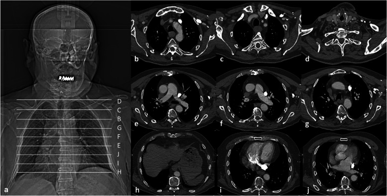
Background: Computed tomography angiography (CTA) of the head and neck during acute ischemic stroke (AIS) usually includes visualization of lung apices. The possibility to evaluate for pulmonary changes, e.g. peripheral ground-glass and consolidative opacities suggestive of coronavirus disease 2019 (COVID-19)-related pneumonia, depends on the area of the lung covered by CTA.
Methods: We performed an analysis of a real-world scenario assessing the variability of lung coverage on CTA in patients presenting with AIS to a comprehensive stroke center (CSC) or to one of eight primary stroke centers (PSC) within a teleradiological network covered by the comprehensive stroke center in 2019.
Results: Our final analysis included n = 940 CTA, and in n = 573 (61%) merely lung apices were covered. In 19/940 (2%) of patients no lung tissue was covered by CTA. CTA scanning protocols in the CSC began significantly more frequently at the level of the ascending aorta (CSC: n = 180 (38.2%), PSC: n = 127 (27.1%), p-value < 0.001) and the aortic arch (CSC: n = 140 (29.7%), PSC: n = 83 (17.7%), p-value < 0.001), and by this covered less frequently the lower lobes compared to CTA acquired in one of the PSC.
Conclusions: In our pre-COVID-19 pandemic representative stroke patient cohort, CTA for AIS covered most often only lung apices. In 37% of the patients CTA visualized at least parts of the lower lobes, the lingula or the middle lobe allowing for a more extensive assessment of the lungs.
Keywords: COVID-19; Computed tomography angiography; Lung; Stroke; Thrombectomy.
Conflict of interest statement
Dr. Pfaff reports personal fees from Stryker outside the submitted work. Ms. Füssel, Mr. Harlan and Dr. Hubert have nothing to disclose. Dr. Bendszus reports personal fees from Boehringer Ingelheim, BBraun, Vascular Dynamics, Bayer, Merck, Teva, Grifols, Springer, grants and personal fees from Novartis and Guerbet, grants from Siemens, Hopp Foundation, from DFG, European Union, Stryker, outside the submitted work.
Figures
Similar articles
-
Variability of acquisition phase of computed tomography angiography in acute ischemic stroke in a real-world scenario.Eur Radiol. 2022 Jan;32(1):281-289. doi: 10.1007/s00330-021-08084-5. Epub 2021 Jun 15. Eur Radiol. 2022. PMID: 34129068 Free PMC article.
-
CTA-for-All: Impact of Emergency Computed Tomographic Angiography for All Patients With Stroke Presenting Within 24 Hours of Onset.Stroke. 2020 Jan;51(1):331-334. doi: 10.1161/STROKEAHA.119.027356. Epub 2019 Nov 5. Stroke. 2020. PMID: 31684848
-
Single-phase CT angiography predicts ASPECTS decay and may help determine when to repeat CT before thrombectomy.J Stroke Cerebrovasc Dis. 2022 Dec;31(12):106815. doi: 10.1016/j.jstrokecerebrovasdis.2022.106815. Epub 2022 Oct 4. J Stroke Cerebrovasc Dis. 2022. PMID: 36206630
-
Incidental COVID-19 related lung apical findings on stroke CTA during the COVID-19 pandemic.J Neurointerv Surg. 2020 Jul;12(7):669-672. doi: 10.1136/neurintsurg-2020-016188. Epub 2020 May 19. J Neurointerv Surg. 2020. PMID: 32430481
-
Ventilation-perfusion lung scanning and spiral computed tomography of the lungs: competing or complementary modalities?Eur J Nucl Med. 1996 Nov;23(11):1547-53. doi: 10.1007/BF01254484. Eur J Nucl Med. 1996. PMID: 8854857 Review.
Cited by
-
Variability of acquisition phase of computed tomography angiography in acute ischemic stroke in a real-world scenario.Eur Radiol. 2022 Jan;32(1):281-289. doi: 10.1007/s00330-021-08084-5. Epub 2021 Jun 15. Eur Radiol. 2022. PMID: 34129068 Free PMC article.
References
-
- Powers WJ, Rabinstein AA, Ackerson T, Adeoye OM, Bambakidis NC, Becker K, et al. Guidelines for the early Management of Patients with acute ischemic stroke: 2019 update to the 2018 guidelines for the early Management of Acute Ischemic Stroke: A guideline for healthcare professionals from the American Heart Association/American Stroke Association. Stroke. 2019;50(12):e344–e418. doi: 10.1161/STR.0000000000000211. - DOI - PubMed
-
- Mayer, S. A., Viarasilpa, T., Panyavachiraporn, N., Brady, M., Scozzari, D., Van Harn, M., Miller, D., Katramados, A., Hefzy, H., Malik, S., Marin, H., Kole, M., Chebl, A., Lewandowski C, Mitsias PD. (2020) CTA-for-All: Impact of Emergency Computed Tomographic Angiography for All Patients With Stroke Presenting Within 24 Hours of Onset. Stroke, 51(1), 331–334. 10.1161/STROKEAHA.119.027356. - PubMed
-
- Esenwa, C., Lee, J.-A., Nisar, T., Shmukler, A., Goldman, I., Zampolin, R., Hsu, K., Labovitz, D., Altschul, D., Haramati, LB. (2020) Utility of apical lung assessment on computed tomography angiography as a COVID-19 screen in acute stroke. Stroke, 51(12), 3765–3769. 10.1161/STROKEAHA.120.030959. - PMC - PubMed
LinkOut - more resources
Full Text Sources
Other Literature Sources