. 2021 Mar 1;6(57):eabd5515.
doi: 10.1126/sciimmunol.abd5515.

Bispecific antibodies targeting mutant RAS neoantigens

Affiliations

Bispecific antibodies targeting mutant RAS neoantigens

Jacqueline Douglass et al. Sci Immunol. .

Abstract

Mutations in the RAS oncogenes occur in multiple cancers, and ways to target these mutations has been the subject of intense research for decades. Most of these efforts are focused on conventional small-molecule drugs rather than antibody-based therapies because the RAS proteins are intracellular. Peptides derived from recurrent RAS mutations, G12V and Q61H/L/R, are presented on cancer cells in the context of two common human leukocyte antigen (HLA) alleles, HLA-A3 and HLA-A1, respectively. Using phage display, we isolated single-chain variable fragments (scFvs) specific for each of these mutant peptide-HLA complexes. The scFvs did not recognize the peptides derived from the wild-type form of RAS proteins or other related peptides. We then sought to develop an immunotherapeutic agent that was capable of killing cells presenting very low levels of these RAS-derived peptide-HLA complexes. Among many variations of bispecific antibodies tested, one particular format, the single-chain diabody (scDb), exhibited superior reactivity to cells expressing low levels of neoantigens. We converted the scFvs to this scDb format and demonstrated that they were capable of inducing T cell activation and killing of target cancer cells expressing endogenous levels of the mutant RAS proteins and cognate HLA alleles. CRISPR-mediated alterations of the HLA and RAS genes provided strong genetic evidence for the specificity of the scDbs. Thus, this approach could be applied to other common oncogenic mutations that are difficult to target by conventional means, allowing for more specific anticancer therapeutics.


Conflict of interest statement

Competing interests: The Johns Hopkins University has filed patent applications related to technologies described in this paper, on which J.D., E.H.-C.H., K.M.W., Q.W., A.D.S., M.S.H., B.J.M., A.H.P., N.P., K.W.K., S.B.G., B.V., and S.Z. are listed as inventors: HLA-restricted epitopes encoded by somatically mutated genes (US20180086832A1), MANAbodies and methods of using (US20200079854A1), MANAbodies targeting tumor antigens and methods of using (PCT/US2020/065617). B.V., K.W.K., and N.P. are founders of Thrive Earlier Detection. K.W.K. and N.P. are consultants to and were on the Board of Directors of Thrive Earlier Detection. B.V., K.W.K., N.P. and S.Z. own equity in Exact Sciences. B.V., K.W.K., N.P., S.Z., and D.M.P. are founders of, hold or may hold equity in, and serve or may serve as consultants to ManaT Bio. B.V., K.W.K., N.P. and S.Z. are founders of, hold equity in, and serve as consultants to Personal Genome Diagnostics. S.Z. has a research agreement with BioMed Valley Discoveries Inc. K.W.K. and B.V. are consultants to Sysmex, Eisai, and CAGE Pharma and hold equity in CAGE Pharma. B.V. is also a consultant to Catalio. K.W.K., B.V., S.Z., and N.P. are consultants to and hold equity in NeoPhore. N.P. is an advisor to and holds equity in CAGE Pharma. C.B. is a consultant to Depuy-Synthes and Bionaut Labs. The companies named above, as well as other companies, have licensed previously described technologies related to this paper from Johns Hopkins University. B.V., K.W.K., S.Z., N.P., and C.B. are inventors on some of these technologies. Licenses to these technologies are or will be associated with equity or royalty payments to the inventors as well as to Johns Hopkins University. The terms of all these arrangements are being managed by Johns Hopkins University in accordance with its conflict of interest policies. Q.W. is the founder and CEO of Complete Omics Inc. M.F.K. received personal fees from Bristol-Myers Squibb and Celltrion. D.M.P. reports grant and patent royalties through institution from BMS, grant from Compugen, stock from Trieza Therapeutics and Dracen Pharmaceuticals, and founder equity from Potenza; being a consultant for Aduro Biotech, Amgen, Astra Zeneca (Medimmune/Amplimmune), Bayer, DNAtrix, Dynavax Technologies Corporation, Ervaxx, FLX Bio, Rock Springs Capital, Janssen, Merck, Tizona, and Immunomic-Therapeutics; being on the scientific advisory board of Five Prime Therapeutics, Camden Nexus II, and WindMil; and being on the board of directors for Dracen Pharmaceuticals. S.B.G. is a founder and holds equity in Advanced Molecular Sciences LLC.

Figures

Fig. 1.
Fig. 1.. Characterization of RAS MANA scFvs.
(A and C to E) Biotinylated G12V or G12WT pHLA-A3 (A) or Q61WT, Q61H, Q61L, or Q61R pHLA-A1 (C to E) was coated on a streptavidin plate at the specified concentrations. Recombinant RAS G12V clone V2 (A), Q61H clone H1 (C), Q61L clone L2 (D), or Q61R clone R6 (E) scFv was incubated in the wells at 1 μg/ml before detection with protein L. ELISAs were performed in duplicate. OD450, optical density at 450 nm. (B and F) T2A3 or SigM5 cells were pulsed with the specified peptides at 50 μM, followed by flow cytometric analysis. (B) T2A3 cells were stained with V2 scFv preconjugated to anti-FLAG-PE, with mean fluorescence intensities (MFIs) plotted. (F) SigM5 cells were stained with clone H1, L2, or R6 phage.
Fig. 2.
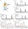
Fig. 2.. Schematic and characterization of MANA scDbs.
(A) Schematic showing the optimal bispecific format, a scDb with variable light (VL or L), and variable heavy (VH or H) domains arranged in the following order: VLV2-VHUCHT1-VLUCHT1-VHV2. SL, short linker (GGGGS); LL, long linker (GGGGS)3. (B and D to F) Anti-MANA/anti-CD3 scDbs were characterized via ELISA. Biotinylated pHLA-A3, pHLA-A1, or CD3ε/δ was coated on a streptavidin plate. V2-U (B), H1-U (D), L2-U (E), or R6-U (F) scDb was incubated at the specified concentrations then detected with protein L. All ELISAs were performed in triplicate. (C and G) V2-U and L2-U scDb binding was evaluated by single-cycle kinetics using SPR. CMV, cytomegalovirus.
Fig. 3.
Fig. 3.. MANA concentration-dependent T cell activation.
(A and B) T2A3 cells were pulsed with either the G12V or G12WT peptides at the specified concentrations. (C and D) SigM5 cells were pulsed with either the Q61L or Q61WT peptides at the specified concentrations. T2A3 (5 × 10⁴) or SigM5 (2.5 × 10⁴) peptide-pulsed cells were combined with 5 × 10⁴ human T cells (effector:target ratio or E:T = 1:1 or 2:1) and V2-U (A and B), V2-U2 scDb (A and B), or L2-U scDb (C and D) at 1 nM. Supernatants were assayed for IFN-γ at 24 hours (A and C) with N = 3 replicates per condition. Target cell cytotoxicity was assayed (B and D) with N = 3 and N = 2 replicates for T2A3 and SigM5 conditions, respectively.
Fig. 4.
Fig. 4.. T cell activation mediated by scDbs recognizing pHLAs formed through endogenous processing.
COS-7 cells were transfected with plasmids encoding HLA-A3 (“A3”) or HLA-A1 (“A1”) and RAS variants or other negative controls. Twenty-four hours later, 1 × 10⁴ COS-7 cells were combined with 5 × 10⁴ T cells (E:T = 5:1) and V2-U (A), H1-U (B), L2-U (C), or R6-U (D) scDb at specific concentrations. Supernatants were assayed for IFN-γ at 24 hours. All experiments were performed in triplicate. GFP, green fluorescent protein.
Fig. 5.
Fig. 5.. V2-U scDb–mediated T cell activation in response to endogenous levels of KRAS G12V pHLA-A3.
Target cells (2 × 10⁴) from parental NCI-H441 (A and B), NCI-H441 variants (C and D), or NCI-H358 variants (E and F) were combined with 6 × 10⁴ T cells (E:T = 3:1) and V2-U scDbs at the specified concentrations. Cells were assayed for IFN-γ release (A, C, and E) and target cell cytotoxicity at 24 hours (B, D, and F). All experiments were performed in triplicate. For KRAS genotypes: G12V/Δ, G12V/frameshift; also see fig. S11.
Fig. 6.
Fig. 6.. L2-U scDb–mediated T cell activation in response to endogenous levels of RAS Q61L pHLA-A1.
Target cells (2.5 × 10⁴) from different cancer cell lines expressing HLA-A1, RAS Q61L, or both (A); parental HL-60 cells (B and C); or HL-60 variants expressing different RAS Q61 mutations or with HLA-A1 KO (D and E) were combined with 5 × 10⁴ T cells (E:T = 2:1) and L2-U scDb at the specified concentrations. Cells were assayed for IFN-γ release by ELISA (A, B, and D) and target cell cytotoxicity at 24 hours (C and E). Experiments were performed with N = 3 (A), N = 4 (B and C), and N = 2 (D and E) replicates per condition. For HL-60 NRAS genotypes, also see fig. S11.
Fig. 7.
Fig. 7.. Peptide scanning mutagenesis to assess potential V2-U and L2-U scDb cross-reactivity.
Each amino acid of the G12V and Q61L peptides was systematically substituted with the other common 19 amino acids, thereby generating libraries of variant peptides each differing from the original peptide by a single amino acid. T2A3 cells were pulsed with 10 μM of individual peptides from the G12V peptide library (A and C), and SigM5 cell cells were pulsed with 10 μM of individual peptides from the Q61L peptide library (B and D). Peptide-pulsed target cells (2.5 × 10⁴) were combined with 5 × 10⁴ human T cells (E:T = 2:1) and either the V2-U (A and C) or L2-U scDb (B and D) at 1 nM. (A and B) Supernatant was assayed for IFN-γ at 24 hours, with the mean of three technical replicates plotted as a heatmap. Black boxes indicate amino acids in the parental peptides. (C and D) Illustration of the binding patterns of V2-U and L2-U scDbs as Seq2Logo graphs, calculated by dividing the IFN-γ value by 103 and using the PSSM-Logo algorithm.

Comment in

References

    1. Vogelstein B, Papadopoulos N, Velculescu VE, Zhou S, Diaz LA Jr., Kinzler KW, Cancer genome landscapes. Science 339, 1546–1558 (2013). - PMC - PubMed
    1. Bedard PL, Hyman DM, Davids MS, Siu LL, Small molecules, big impact: 20 years of targeted therapy in oncology. Lancet 395, 1078–1088 (2020). - PubMed
    1. Wang QJ, Yu Z, Griffith K, Hanada K, Restifo NP, Yang JC, Identification of T-cell receptors targeting KRAS-mutated human tumors. Cancer Immunol. Res 4, 204–214 (2016). - PMC - PubMed
    1. Canon J, Rex K, Saiki AY, Mohr C, Cooke K, Bagal D, Gaida K, Holt T, Knutson CG, Koppada N, Lanman BA, Werner J, Rapaport AS, Miguel TS, Ortiz R, Osgood T, Sun JR, Zhu X, McCarter JD, Volak LP, Houk BE, Fakih MG, O’Neil BH, Price TJ, Falchook GS, Desai J, Kuo J, Govindan R, Hong DS, Ouyang W, Henary H, Arvedson T, Cee VJ, Lipford JR, The clinical KRAS(G12C) inhibitor AMG 510 drives anti-tumour immunity. Nature 575, 217–223 (2019). - PubMed
    1. Tran E, Robbins PF, Lu YC, Prickett TD, Gartner JJ, Jia L, Pasetto A, Zheng Z, Ray S, Groh EM, Kriley IR, Rosenberg SA, T-cell transfer therapy targeting mutant KRAS in cancer. N. Engl. J. Med 375, 2255–2262 (2016). - PMC - PubMed

Publication types

MeSH terms