. 2021 Apr 21;203(10):e00599-20.
doi: 10.1128/JB.00599-20. Print 2021 Apr 21.

Evolution of Ribosomal Protein S14 Demonstrated by the Reconstruction of Chimeric Ribosomes in Bacillus subtilis

Affiliations

Evolution of Ribosomal Protein S14 Demonstrated by the Reconstruction of Chimeric Ribosomes in Bacillus subtilis

Genki Akanuma et al. J Bacteriol. .

Abstract

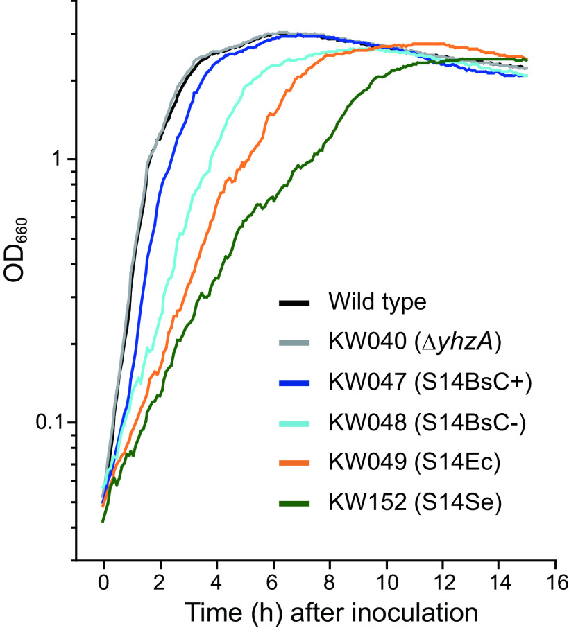
Ribosomal protein S14 can be classified into three types. The first, the C+ type has a Zn2+ binding motif and is ancestral. The second and third are the C- short and C- long types, neither of which contain a Zn2+ binding motif and which are ca. 90 residues and 100 residues in length, respectively. In the present study, the C+ type S14 from Bacillus subtilis ribosomes (S14BsC+) were completely replaced by the heterologous C- long type of S14 from Escherichia coli (S14Ec) or Synechococcus elongatus (S14Se). Surprisingly, S14Ec and S14Se were incorporated fully into 70S ribosomes in B. subtilis However, the growth rates as well as the sporulation efficiency of the mutants harboring heterologous S14 were significantly decreased. In these mutants, the polysome fraction was decreased and the 30S and 50S subunits accumulated unusually, indicating that cellular translational activity of these mutants was decreased. In vitro analysis showed a reduction in the translational activity of the 70S ribosome fraction purified from these mutants. The abundance of ribosomal proteins S2 and S3 in the 30S fraction in these mutants was reduced while that of S14 was not significantly decreased. It seems likely that binding of heterologous S14 changes the structure of the 30S subunit, which causes a decrease in the assembly efficiency of S2 and S3, which are located near the binding site of S14. Moreover, we found that S3 from S. elongatus cannot function in B. subtilis unless S14Se is present.IMPORTANCE S14, an essential ribosomal protein, may have evolved to adapt bacteria to zinc-limited environments by replacement of a zinc-binding motif with a zinc-independent sequence. It was expected that the bacterial ribosome would be tolerant to replacement of S14 because of the previous prediction that the spread of C- type S14 involved horizontal gene transfer. In this study, we completely replaced the C+ type of S14 in B. subtilis ribosome with the heterologous C- long type of S14 and characterized the resulting chimeric ribosomes. Our results suggest that the B. subtilis ribosome is permissive for the replacement of S14, but coevolution of S3 might be required to utilize the C- long type of S14 more effectively.

Keywords: Bacillus subtilis; ribosomal protein S14; ribosome; zinc.


Figures

FIG 1
FIG 1
Phylogenetic tree and alignment of ribosomal protein S14. (A) Protein sequences, available from the NCBI database (https://www.ncbi.nlm.nih.gov/protein), were aligned, and a phylogenetic tree was constructed by the maximum likelihood method and Le-Gascuel model (59) with the use of MEGA X software (52). Bootstrapping was performed with 1,000 replicates. The scale bar (0.50) indicates the number of changes per site. “[B]” after a species name indicates that the species have both types (C+ and C−) of S14. (B) Alignment of the amino acid sequences of S14 from B. subtilis, E. coli, and S. elongatus. Identical and similar amino acids are highlighted with yellow and blue, respectively. Cysteines forming the zinc-binding motif and remnants of this motif in sequences that do not have all four conserved cysteines are shown in bold.
FIG 2
FIG 2
Effect of the replacement of S14BsC+ by heterologous S14 on the growth rate. Cells were grown in LB at 37°C, and the optical density at 660 nm was measured.
FIG 3
FIG 3
Component proteins of 70S ribosomes prepared from cells containing heterologous S14. The upper panel shows whole 2D gel of ribosomal proteins from 70S ribosomes from wild type. The areas of the two-dimensional gels containing the spot corresponding to each S14 were extracted from gel images (lower panels). Spots corresponding to S14 are indicated by closed arrowheads, and the absence of the protein spot corresponding to S14Bs upon replacement is indicated by open arrowheads.
FIG 4
FIG 4
Decrease in the polysome fraction and accumulation of the 30S and 50S subunits in cells harboring S14Ec or S14Se. Crude cell extracts were sedimented through a 10 to 40% sucrose gradient as described in Materials and Methods. The 30S, 50S, 70S, and polysome peaks are indicated in each individual profile. Abs, absorbance.
FIG 5
FIG 5
In vitro translation analysis using 70S ribosomes containing heterologous S14. The relative amount of GFP, which was synthesized by each 70S ribosome, is shown as relative activity where the amount of GFP synthesized by wild-type 70S is defined as 1. The 70S ribosomes were purified from wild-type, KW047 (S14BsC+), KW048 (S14BsC−), KW049 (S14Ec), and KW152 (S14Se) cells.
FIG 6
FIG 6
Effect of the replacement of S14BsC+ by heterologous S14 on the assembly of the 30S subunit. (A) Two-dimensional electrophoresis analysis of the component proteins of each 30S subunit prepared from cells containing heterologous S14. Ribosomal protein spots are indicated by closed arrowheads, and the absence of the protein spot corresponding to S14Bs by replacement is indicated by open arrowheads. (B) Comparison of ribbon diagram of S14BsC+ (cyan) with that of S14BsC− (yellow), S14Ec (green), and S14Se (magenta). The cysteine residues that form the zinc-binding motif of S14BsC+ are highlighted in red. The protein structures of S14BsC+, S14BsC−, S14Ec, and S14Se were predicted by SWISS-MODEL (http://swissmodel.expasy.org/) (60) using the solved structure of B. subtilis ribosome (Protein Data Bank [PDB] accession number 5NJT), Mycobacterium smegmatis C− ribosome (PDB accession number 6DZI), E. coli ribosome (PDB accession number 3J9Y), and chloroplast ribosome of Spinacia oleracea (PDB accession number 6ERI) as the template. An image of the resulting structure was generated using PyMOL (www.pymol.org) (61). The N-terminal extrusion is highlighted with a dotted circle (see details in text). (C) Location of S2 (cyan), S3 (green), and S14 (red) in the B. subtilis 70S ribosome are shown using the solved structure (PDB accession number 5NJT). The 16S rRNA in the 30S subunit is shown in blue.
FIG 7
FIG 7
The replacement of S14BsC+ by heterologous S14 causes a sporulation defect. (A) Microscopic observation of wild-type and mutants harboring S14Ec or S14Se cells. Differential interference contrast and fluorescence images of FM4-64 stained (to visualize membranes) and DAPI stained (to visualize chromosomes) cells grown for indicated times at 37°C in sporulation medium. The arrowheads in the micrographs indicate prespores. Bars indicate 10 μm. (B) Cells were grown in sporulation medium at 37°C and were collected at the indicated times. Crude cell extracts were subjected to Western blot analysis using antisera against Spo0A. (C) Cells were grown in sporulation medium at 37°C, and the optical density at 600 nm was measured.
FIG 8
FIG 8
The replacement of S3Bs by S3Se in the presence of S14BsC+ and/or S14Se. (A) Strains KW158 (presence of S14Bs and S14Se) and KW159 (presence of only S14Se) were streaked onto an LB plate without (left plate) or with (right plate) 1 mM IPTG. KW158 grew only when S3Bs and S2Bs were expressed, whereas KW159 did not require the induction of S3Bs expression. (B) Cells grown on the LB plate containing 1 mM IPTG were inoculated into LB medium without or with 1 mM IPTG and cultured at 37°C, and the optical density at 600 nm was measured.

Similar articles

Cited by

References

    1. Kurland CG. 1972. Structure and function of the bacterial ribosome. Annu Rev Biochem 41:377–408. 10.1146/annurev.bi.41.070172.002113. - DOI - PubMed
    1. Nomura M. 1970. Bacterial ribosome. Bacteriol Rev 34:228–277. 10.1128/BR.34.3.228-277.1970. - DOI - PMC - PubMed
    1. Gao YG, Selmer M, Dunham CM, Weixlbaumer A, Kelley AC, Ramakrishnan V. 2009. The structure of the ribosome with elongation factor G trapped in the posttranslocational state. Science 326:694–699. 10.1126/science.1179709. - DOI - PMC - PubMed
    1. Schmeing TM, Ramakrishnan V. 2009. What recent ribosome structures have revealed about the mechanism of translation. Nature 461:1234–1242. 10.1038/nature08403. - DOI - PubMed
    1. Schmeing TM, Voorhees RM, Kelley AC, Gao YG, Murphy FV, Weir JR, Ramakrishnan V. 2009. The crystal structure of the ribosome bound to EF-Tu and aminoacyl-tRNA. Science 326:688–694. 10.1126/science.1179700. - DOI - PMC - PubMed

Publication types

MeSH terms

Supplementary concepts

LinkOut - more resources