Review
. 2021 Mar 1;35(5-6):307-328.
doi: 10.1101/gad.346312.120.

Chronic tissue inflammation and metabolic disease

Affiliations
Review

Chronic tissue inflammation and metabolic disease

Yun Sok Lee et al. Genes Dev. .

Abstract

Obesity is the most common cause of insulin resistance, and the current obesity epidemic is driving a parallel rise in the incidence of T2DM. It is now widely recognized that chronic, subacute tissue inflammation is a major etiologic component of the pathogenesis of insulin resistance and metabolic dysfunction in obesity. Here, we summarize recent advances in our understanding of immunometabolism. We discuss the characteristics of chronic inflammation in the major metabolic tissues and how obesity triggers these events, including a focus on the role of adipose tissue hypoxia and macrophage-derived exosomes. Last, we also review current and potential new therapeutic strategies based on immunomodulation.

Keywords: glucose intolerance; immunometabolism; inflammation; insulin resistance; macrophage; metaflammation; β-cell dysfunction.


Figures

Figure 1.
Figure 1.
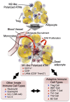
Adipose tissue inflammation in obesity. In the normal state, resident ATMs mostly show an M2-like polarized phenotype. Factors released from Tregs and eosinophils support ATMs to maintain this anti-inflammatory state. In obesity, increased adipocyte chemokine production induces increased blood monocyte recruitment, as well as ATM proliferation. The majority of monocyte-derived ATMs express CD11c and/or CD9 and the M1-like polarized phenotype. The decreased number of eosinophils and Tregs and the increased number of neutrophils, ILC1, CD8+ T cells, Th1 cells, and B2 cells enhance M1-like ATM polarization and adipose tissue inflammation.
Figure 2.
Figure 2.
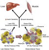
Inflammation in the liver. In the normal physiological state, KCs account for ∼10% of all liver cells. In addition, they scavenge pathogens and show M2-like polarized anti-inflammatory phenotype. In obese steatotic livers, KCs can show increased expression of genes associated with tissue repair and inflammation. These genes include Cd9 and Trem2. This is accompanied by increased KC apoptosis, and a component of KC death is compensated for by increased recruitment of blood monocytes, which differentiate into KC-like macrophages. Chemokines released by steatotic hepatocytes cause increased recruitment of blood monocytes into the liver, which differentiate into proinflammatory macrophages (RHMs) and produce factors that can cause insulin resistance. The recruitment of neutrophils also increases, and the molecules released from neutrophil granules such as neutrophil elastase and myeloperoxidase can induce insulin resistance in hepatocytes.
Figure 3.
Figure 3.
Intracellular adipocyte hypoxia triggers inflammation and insulin resistance. In obesity, increased intracellular FFAs stimulate ANT2-dependent increased mitochondrial uncoupled respiration in adipocytes. Combined with decreased functional capillary density, this leads to intracellular hypoxia and HIF-1α stabilization. HIF-1α induces increased chemokine production, leading to increased immune cell infiltration, including monocytes, and increased ATM proliferation. These changes critically affect ATM exosome secretion, as well as adipocyte exosome and adipokine production, causing systemic insulin resistance. Most of these sequential events have been shown in mouse models. This hypothesis remains to be validated in humans.
Figure 4.
Figure 4.
Systemic effect of ATM exosomes. ATMs can regulate systemic insulin sensitivity by releasing miRNA-containing exosomes. Injection of exosomes released from lean mouse ATMs increases insulin sensitivity in obese mice, whereas injection of obese mouse ATM-derived exosomes induces insulin resistance in lean mice. In addition, direct in vitro treatment of insulin target cells with “lean” or “obese” exosomes directly causes insulin sensitivity or resistance, respectively. Most of these sequential events have been shown in mouse models. This hypothesis remains to be validated in humans.

Similar articles

Cited by

References

    1. Alonge KM, D'Alessio DA, Schwartz MW. 2021. Brain control of blood glucose levels: implications for the pathogenesis of type 2 diabetes. Diabetologia 64: 5–14. - PMC - PubMed
    1. Amano SU, Cohen JL, Vangala P, Tencerova M, Nicoloro SM, Yawe JC, Shen Y, Czech MP, Aouadi M. 2014. Local proliferation of macrophages contributes to obesity-associated adipose tissue inflammation. Cell Metab 19: 162–171. 10.1016/j.cmet.2013.11.017 - DOI - PMC - PubMed
    1. Amar S, Zhou Q, Shaik-Dasthagirisaheb Y, Leeman S. 2007. Diet-induced obesity in mice causes changes in immune responses and bone loss manifested by bacterial challenge. Proc Natl Acad Sci 104: 20466–20471. 10.1073/pnas.0710335105 - DOI - PMC - PubMed
    1. Amar J, Chabo C, Waget A, Klopp P, Vachoux C, Bermúdez-Humarán LG, Smirnova N, Bergá M, Sulpice T, Lahtinen S, et al. 2011. Intestinal mucosal adherence and translocation of commensal bacteria at the early onset of type 2 diabetes: molecular mechanisms and probiotic treatment. EMBO Mol Med 3: 559–572. 10.1002/emmm.201100159 - DOI - PMC - PubMed
    1. Ang Z, Ding JL. 2016. GPR41 and GPR43 in obesity and inflammation: protective or causative? Front Immunol 7: 28. - PMC - PubMed

Publication types