. 2021 Mar 9;118(10):e2012171118.
doi: 10.1073/pnas.2012171118.

Structural insights into α-synuclein monomer-fibril interactions

Affiliations

Structural insights into α-synuclein monomer-fibril interactions

Pratibha Kumari et al. Proc Natl Acad Sci U S A. .

Abstract

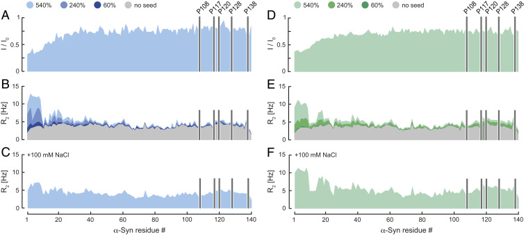
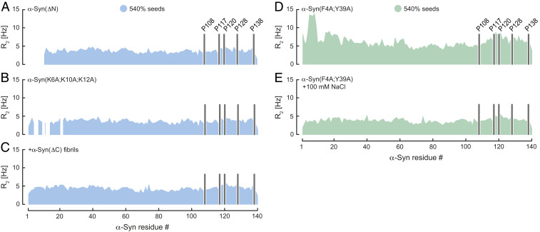
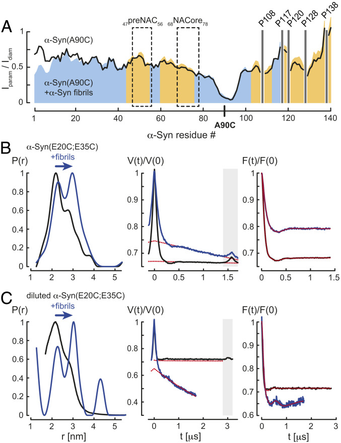
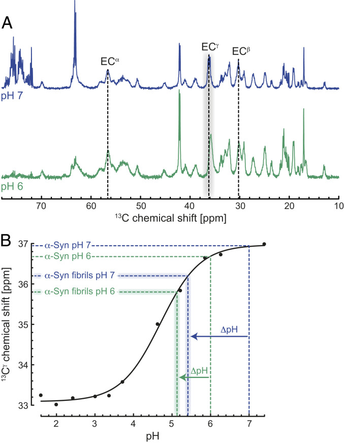
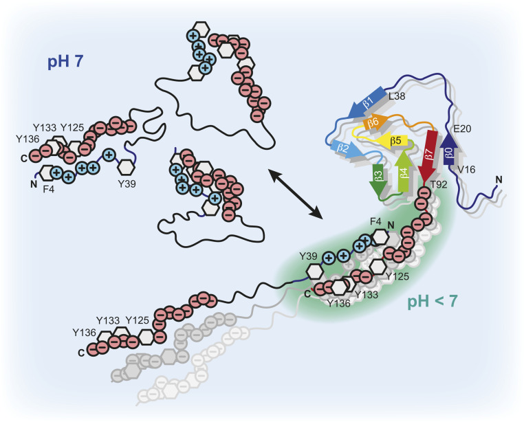
Protein aggregation into amyloid fibrils is associated with multiple neurodegenerative diseases, including Parkinson's disease. Kinetic data and biophysical characterization have shown that the secondary nucleation pathway highly accelerates aggregation via the absorption of monomeric protein on the surface of amyloid fibrils. Here, we used NMR and electron paramagnetic resonance spectroscopy to investigate the interaction of monomeric α-synuclein (α-Syn) with its fibrillar form. We demonstrate that α-Syn monomers interact transiently via their positively charged N terminus with the negatively charged flexible C-terminal ends of the fibrils. These intermolecular interactions reduce intramolecular contacts in monomeric α-Syn, yielding further unfolding of the partially collapsed intrinsically disordered states of α-Syn along with a possible increase in the local concentration of soluble α-Syn and alignment of individual monomers on the fibril surface. Our data indicate that intramolecular unfolding critically contributes to the aggregation kinetics of α-Syn during secondary nucleation.

Keywords: Parkinson’s disease; protein aggregation; secondary nucleation; α-synuclein.


Conflict of interest statement

The authors declare no competing interest.

Figures

Fig. 1.
Fig. 1.
Transient α-Syn monomer–fibril interaction. (A and D) Residue-resolved NMR signal intensity ratios (I/I0) of fibril-bound (I) and unbound (I0) α-Syn at pH 7 (blue) and 6 (green), no salt. 15N R2 relaxation rates of fibril-bound α-Syn in the (B and E) absence and (C and F) presence of 100 mM NaCl at pH 7 and 6. Seed concentrations are expressed as percentage of soluble α-Syn concentration. I/I0 was determined at 540% α-Syn fibril seeds. R2 rates were measured at 0, 60, 240, and 540% α-Syn fibril seeds. Positions of C-terminal α-Syn proline residues without peptide amide resonances are shown in one-letter amino acid code.
Fig. 2.
Fig. 2.
Transient α-Syn mutant monomer–fibril interaction. Residue-resolved 15N R2 relaxation rates of (A) α-Syn(ΔN), (B) α-Syn(K6A;K10A;K12A), and (C) α-Syn in presence of wild-type α-Syn and α-Syn(ΔC) fibrils at pH 7. R2 of α-Syn(F4A;Y39A) upon addition of wild-type α-Syn fibril seeds in the (D) absence and (E) presence of 100 mM NaCl at pH 6. Seed concentration is expressed as percentage of soluble α-Syn concentration. Positions of C-terminal α-Syn proline residues without peptide amide resonances are shown in one-letter amino acid code.
Fig. 3.
Fig. 3.
Unfolding of fibril-bound α-Syn. (A) Residue-resolved PRE intensity profiles Iparam/Idiam of para- (Iparam) and diamagnetic (Idiam) labeled α-Syn(Α90C) in the absence (black) and presence of 5.4-fold molar excess of α-Syn fibrils (blue) at pH 7. Regions with increased Iparam/Idiam values in presence of α-Syn fibrils compared to soluble α-Syn(Α90C) are colored in orange. The preNAC and NACore regions of α-Syn are highlighted. Positions of C-terminal α-Syn proline residues without peptide amide resonances are shown in one-letter amino acid code. (B) EPR measurements of MTSL-labeled α-Syn(E20C;E35C) and (C) partially MTSL-labeled α-Syn(E20C;E35C) in the absence (black) and presence (blue) of α-Syn fibrils, monomer:fibril molar ratio 1:10, at pH 7. From left to right, distance distributions P(r) using DeerAnalysis2019, primary Q-band DEER data V(t)/V(0), and background-corrected form factors F(t)/F(0) are shown including homogeneous background fits (red dotted lines). Time traces within the gray boxes were excluded from the background.
Fig. 4.
Fig. 4.
Lowered local pH on the surface of the α-Syn fibrils triggers monomer–fibril interactions. (A) One-dimensional (1D) 13C MAS INEPT NMR spectra of the mobile regions of α-Syn fibrils at pH 7 (blue) and 6 (green) in the absence of salt. 13C chemical shifts of Glu residues are shown in one-letter amino acid code. (B) Comparison of the pH titration data reported for Glu114 13Cγ of soluble α-Syn (black, reproduced from ref. 39) and the Glu 13Cγ chemical shifts of the α-Syn fibrils obtained from the 1D 13C INEPT spectrum. The local ΔpH differences between soluble and α-Syn fibrils at pH 7 (blue) and 6 (green) are indicated.
Fig. 5.
Fig. 5.
Scheme of α-Syn fibril interactions. Transient long-range intramolecular contacts between N- and C-terminal residues in soluble α-Syn yield partially collapsed intrinsically disordered states of the protein and occlude the central aggregation-prone NAC region. The highly negatively charged C terminus of the α-Syn fibrils lowers the local pH compared to the bulk. Intermolecular α-Syn-fibril interactions between the positively charged N-terminal segment of α-Syn and the negatively charged flexible C-terminal tails of the fibrils unfold the loosely packed α-Syn structures and dynamically align α-Syn molecules at high local concentrations on the fibril surface. This results in an exposure of the NAC region and triggers protein aggregation via secondary nucleation.

References

    1. Spillantini M. G., et al. ., Alpha-synuclein in Lewy bodies. Nature 388, 839–840 (1997). - PubMed
    1. Spillantini M. G., Goedert M., The α-synucleinopathies: Parkinson’s disease, dementia with Lewy bodies, and multiple system atrophy. Ann. N. Y. Acad. Sci. 920, 16–27 (2000). - PubMed
    1. Krüger R., et al. ., Ala30Pro mutation in the gene encoding alpha-synuclein in Parkinson’s disease. Nat. Genet. 18, 106–108 (1998). - PubMed
    1. Zarranz J. J., et al. ., The new mutation, E46K, of alpha-synuclein causes Parkinson and Lewy body dementia. Ann. Neurol. 55, 164–173 (2004). - PubMed
    1. Appel-Cresswell S., et al. ., Alpha-synuclein p.H50Q, a novel pathogenic mutation for Parkinson’s disease. Mov. Disord. 28, 811–813 (2013). - PubMed

Publication types

LinkOut - more resources