. 2021 Mar 1;12(1):1362.
doi: 10.1038/s41467-021-21630-5.

Chemogenetic ON and OFF switches for RNA virus replication

Affiliations

Chemogenetic ON and OFF switches for RNA virus replication

E Heilmann et al. Nat Commun. .

Abstract

Therapeutic application of RNA viruses as oncolytic agents or gene vectors requires a tight control of virus activity if toxicity is a concern. Here we present a regulator switch for RNA viruses using a conditional protease approach, in which the function of at least one viral protein essential for transcription and replication is linked to autocatalytical, exogenous human immunodeficiency virus (HIV) protease activity. Virus activity can be en- or disabled by various HIV protease inhibitors. Incorporating the HIV protease dimer in the genome of vesicular stomatitis virus (VSV) into the open reading frame of either the P- or L-protein resulted in an ON switch. Here, virus activity depends on co-application of protease inhibitor in a dose-dependent manner. Conversely, an N-terminal VSV polymerase tag with the HIV protease dimer constitutes an OFF switch, as application of protease inhibitor stops virus activity. This technology may also be applicable to other potentially therapeutic RNA viruses.


Conflict of interest statement

A patent application relating to all aspects of the manuscript has been filed under the application number 19181717 by Boehringer Ingelheim International GmbH (application date 21 June 2019). E.H., J.K., B.H., L.E., T.N., G.W., and D.v.L. are listed as inventors. T.N., C.U., and L.E. are employed by ViratTherapeutics GmbH. D.v.L. is founder of ViraTherapeutics GmbH. D.v.L. and G.W. serve as scientific advisors to Boehringer Ingelheim Pharma K.G. ViraTherapeutics or Boehringer Ingelheim had no role in the study design, data analysis and interpretation, or the writing of the manuscript. The other authors declare no competing interests.

Figures

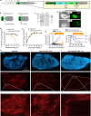
Fig. 1
Fig. 1. VSV-Pprot and VSV-Lprot require the presence of HIV protease inhibitor for virus activity.
a Scheme of the VSV genome with detailed depiction of the intramolecular insertion into the phosphoprotein (P) of the HIV protease linked-dimer construct flanked by protease cleavage sites and GGSG linker sequences. b Model of the intact VSV nucleoprotein-phosphoprotein-polymerase (N-P-L) replication complex in the presence of protease inhibitor. Absence of protease inhibitor results in autoproteolytic separation of P into N- and C-terminal fragments (PN′, PC′) and disengagement from the replication complex. c Western Blot against HIV protease display the large fusion protein in presence of protease inhibitor (+PI) resolved prior on low percentage polyacrylamide gel. Protein samples without protease inhibitor (-PI) were produced on replication-supporting 293-VSV cells expressing N, P, and L and resolved on a high percentage polyacrylamide gel. d Scheme of the intramolecular insertion into the VSV L protein of the same protease dimer construct. e Model of the intact VSV L protein in the presence of protease inhibitor. Absence of protease inhibitor results in autoproteolytic separation of L into two fragments (LN′, LC′) and halted polymerase activity. f Western Blot against HIV protease with protease inhibitor (+PI) from large-pore gel and without protease inhibitor (-PI) from small-pore gel. Virus replication on BHK cells leading to GFP viral reporter gene expression (g) required the presence of PI amprenavir (APV) in a dose-dependent manner for VSV-Pprot-GFP (h) and VSV-Lprot-GFP (i). BHK cells were infected with VSV-GFP, VSV-Pprot-GFP, or VSV-Lprot-GFP at an MOI of 1 in the presence of increasing doses of APV. Supernatant was collected 24 h later and virus progeny titer was determined via TCID50 assay (technical replicates; n = 2). j Replication kinetic of VSV-GFP vs. VSV-Lprot-GFP and VSV-Pprot-GFP. BHK cells were infected with VSV-GFP, VSV-Lprot-GFP, or VSV-Pprot-GFP at an MOI of 3 in the presence of 10 µM APV. Supernatants were collected at indicated time points and virus progeny titer was determined via TCID50 assay (technical replicates; n = 3; dotted line indicates detection limit). Western blot experiments in panels (c, f) were performed three times. Viral growth curve studies in panels (h, i, j) were performed two times times. Source data are provided in the Source Data File.
Fig. 2
Fig. 2. VSV-Pprot and VSV-Lprot are regulatable in vivo.
a Subcutaneous human glioma U87 xenografts were intratumorally treated with luciferase-expressing VSV-Pprot-Luc with (+PI) or without (-PI) concomitant intraperitoneal (i.p.) application of APV. Representative bioluminescence (BLI) images are shown from 8 days after virus injection. b BLI quantification of luciferase signal from VSV-Pprot-Luc treated tumors in mice receiving PI cocktail (APV + RTV) (red) or drug vehicle (blue) (technical replicates; n = 5; * denotes significantly different measurements with p < 0.05; unpaired two-tailed t test at indicated time points). c, d Single intratumoral treatment of subcutaneus U87 tumors with VSV (n = 6) or VSV-Lprot-GFP with (n = 5) or without (n = 5) PI treatment (APV + RTV) (mock n = 6). Log-rank (Mantel-Cox) test was performed (**p = 0.0052). eh 2 μl of VSV-DsRed or VSV-Pprot-GFP were stereotactically injected into the striatum of BALB/c mice. PI (APV + RTV) treatment was applied every 12 h for 9 days. VSV-Pprot-GFP was well tolerated with no significant weight loss (e) or signs of neurotoxicity (f) compared to fatal neurotoxicity of parental VSV-DsRed (g) (**p = 0.0078; Log-rank (Mantel-Cox) test). Symbols in f display score per mouse per time point). h Histological fluorescence analysis of coronal brain sections revealed extended spread of VSV-DsRed in the striatum, subcortical areas and hypothalamus (bilateral) at 3 days post inoculation (dpi). In contrast, GFP expression from VSV-Pprot-GFP (10 dpi) was restricted to the immediate lining of the injection needle track without any signs of intracranial spread, irrespective of i.p. co-treatment of PI or drug vehicle. PI treatment with high-dose IDV to increase CNS availability after VSV-Lprot-GFP injection did also not induce signs of neurotoxicity and brain parenchymal spread was restricted to the injection site (i, j); n = 3 for VSV-GFP, n = 5 for Lprot variants. Bioluminescence experiments in panels (a, b) and associated tumor growth and survival study (c, d) were performed once. Intracranial injection experiments (eh) were performed two times. Source data are provided in the Source Data File.
Fig. 3
Fig. 3. N-terminal fusion of protease dimer construct to VSV L protein constitutes a regulatable OFF-switch.
a Scheme of the VSV genome with detailed depiction of the N-terminal fusion construct of the linked-dimer protease construct flanked by autoproteolytic cleavage sites with the L protein. b Scheme of the autoproteolytic detachment of the protease dimer construct and GFP from the polymerase (L) in absence of protease inhibitor and generation of fusion protein leading to GFP viral reporter gene expression in infected BHK cells in the absence of PI. c Western Blot against HIV protease display the released HIV protease dimer in the absence of protease inhibitor (-PI) resolved prior on high percentage polyacrylamide gel. Protein samples with protease inhibitor (+PI) were produced on replication-supporting 293-VSV cells expressing N, P, and L and resolved on a low percentage polyacrylamide gel. Treatment with PI (here saquinavir SQV, 10 µM) results in dysfunctional GFP-prot-L fusion. d The GFP-prot-L fusion protein can be visualized by infection of cells with high multiplicity of infection and addition of PI. Incoming virions carry functional L that produces the large fusion protein, which accumulates in typical rhabdovirus replication foci (lower panel). Without PI, GFP fluorescence is diffuse, infected cells round up and die (upper panel). e Dose response of VSV-GFP and VSV-Prot-OFF-GFP for protease inhibitor saquinavir (SQV). BHK cells were infected with VSV-GFP or VSV-Prot-OFF-GFP at an MOI of 1 in the presence of increasing doses of SQV. Supernatant was collected 24 hrs later and virus progeny titer was determined via TCID50 assay (n = 2; technical replicates). f Replication kinetics of VSV-GFP vs. VSV-Prot-OFF-GFP. BHK cells were infected with VSV-GFP or VSV-Prot-OFF-GFP at an MOI of 3. Supernatants were collected at indicated time points and virus progeny titer was determined via TCID50 assay (n = 3; technical replicates). g NOD-SCID mice bearing subcutaneous human glioma G62 xenografts were treated intratumorally twice with 7 day interval (dotted line) with either VSV-GFP (n = 8), VSV-Prot-OFF-GFP without (n = 8) or with (n = 8) PI treatment (SQV + RTV) 3x a day. Orange bar indicates start and duration of PI treatment. Left panel shows individual tumor growth kinetics. Right panel depicts the Kaplan–Meier survival curves (Log-rank (Mantel-Cox) test, p values from comparison to PBS control). In a parallel study, PI (SQV + RTV) treatment was initiated 3 days after single virus injection and tumors treated with VSV-GFP or VSV-Prot-OFF-GFP (n = 3) were harvested 1 week later and analyzed for virus spread using anti-VSV immunofluorescence staining. Representative images show wide intratumoral spread of VSV-GFP (h), disseminated spread of VSV-Prot-OFF-GFP without SQV (i) and isolated reduced virus staining of VSV-Prot-OFF-GFP under SQV treatment (j). Western blot experiments in panel (c) were performed three times. High-resolution fluorescence microscopy experiments in panel d were repeated twice with 2 wells per condition. 10 positions of each well were monitored automatically for up to 18 h post infection. Viral growth curve studies in panels (e, f) were performed two times times. Tumor growth and survival study in panel (g) were performed once. Source data are provided in the Source Data File.

References

    1. Bouard D, Alazard-Dany D, Cosset FL. Viral vectors: from virology to transgene expression. Br. J. Pharmacol. 2009;157:153–165. doi: 10.1038/bjp.2008.349. - DOI - PMC - PubMed
    1. Kaufman HL, Kohlhapp FJ, Zloza A. Oncolytic viruses: a new class of immunotherapy drugs. Nat. Rev. Drug Discov. 2015;14:642–662. doi: 10.1038/nrd4663. - DOI - PMC - PubMed
    1. Henao-Restrepo AM, et al. Efficacy and effectiveness of an rVSV-vectored vaccine expressing Ebola surface glycoprotein: interim results from the Guinea ring vaccination cluster-randomised trial. Lancet. 2015;386:857–866. doi: 10.1016/S0140-6736(15)61117-5. - DOI - PubMed
    1. Meissner HC, Strebel PM, Orenstein WA. Measles vaccines and the potential for worldwide eradication of measles. Pediatrics. 2004;114:1065–1069. doi: 10.1542/peds.2004-0440. - DOI - PubMed
    1. Goverdhana S, et al. Regulatable gene expression systems for gene therapy applications: progress and future challenges. Mol. Ther. 2005;12:189–211. doi: 10.1016/j.ymthe.2005.03.022. - DOI - PMC - PubMed

Publication types