. 2021 Mar 1;12(1):1346.
doi: 10.1038/s41467-021-21634-1.

S-Trimer, a COVID-19 subunit vaccine candidate, induces protective immunity in nonhuman primates

Affiliations

S-Trimer, a COVID-19 subunit vaccine candidate, induces protective immunity in nonhuman primates

Joshua G Liang et al. Nat Commun. .

Abstract

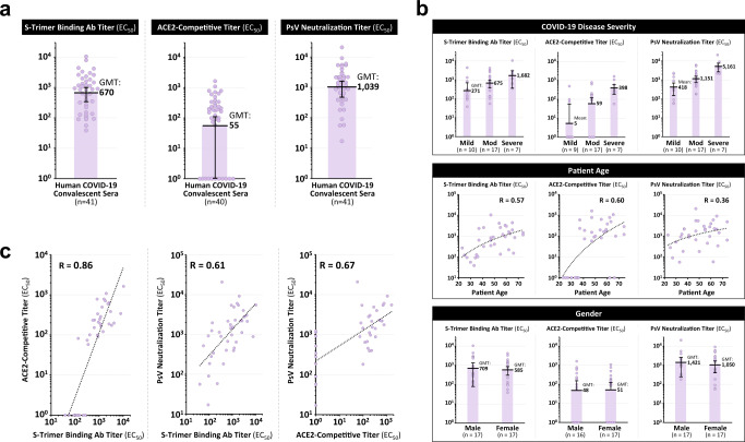
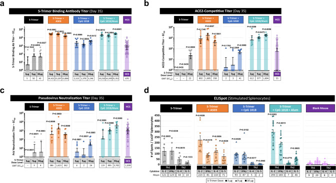
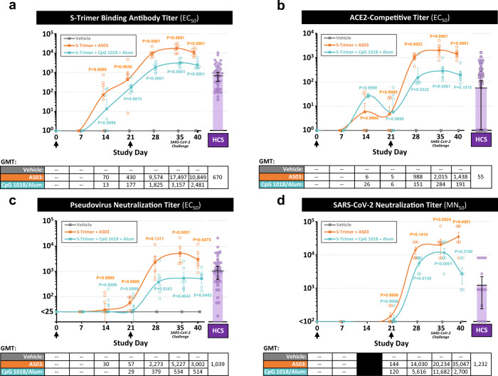
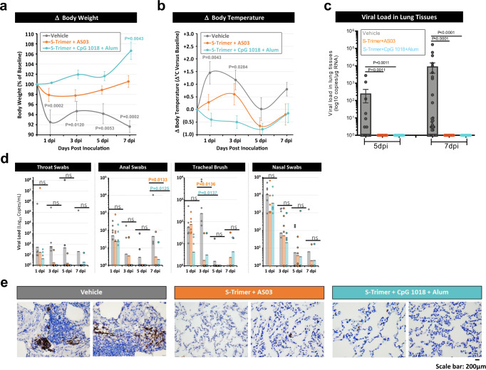
SARS-CoV-2 is the underlying cause for the COVID-19 pandemic. Like most enveloped RNA viruses, SARS-CoV-2 uses a homotrimeric surface antigen to gain entry into host cells. Here we describe S-Trimer, a native-like trimeric subunit vaccine candidate for COVID-19 based on Trimer-Tag technology. Immunization of S-Trimer with either AS03 (oil-in-water emulsion) or CpG 1018 (TLR9 agonist) plus alum adjuvants induced high-level of neutralizing antibodies and Th1-biased cellular immune responses in animal models. Moreover, rhesus macaques immunized with adjuvanted S-Trimer were protected from SARS-CoV-2 challenge compared to vehicle controls, based on clinical observations and reduction of viral loads in lungs. Trimer-Tag may be an important platform technology for scalable production and rapid development of safe and effective subunit vaccines against current and future emerging RNA viruses.


Conflict of interest statement

J.G.L. and P.L. have ownership interest in Clover Biopharmaceuticals. All other authors have no competing interests.

Figures

Fig. 1
Fig. 1. High-level expression and characterization of S-Trimer.
a Schematic representations of full-length SARS-CoV-2 Spike (S) protein and the ectodomain of wild-type SARS-CoV-2 S protein-Trimer-Tag fusion protein (S-Trimer). b Schematic 2-D illustration of S-Trimer with homotrimeric Spike protein in the prefusion conformation. c Reducing SDS-PAGE analysis with Coomassie Blue staining of high-level expression of S-Trimer as a secreted protein from CHO cells in a 15L bioreactor Fed-batch serum-free culture over 11 days (10 µL of cleared media were loaded for each sample) along with a purified standard (Std). d S-Trimer is a disulfide bond-linked homotrimer as analyzed by SDS-PAGE with Coomassie Blue staining under non-reducing (-ME) and reducing (+ME) conditions. S-Trimer was shown to be partially cleaved at S1/S2 junction as indicated. e S-Trimer is heavily N-glycosylated. Analysis of S-Trimer before and after deglycosylation with PNGase F (-N) and PNGase F & Endo-O (-O) by SDS-PAGE with Coomassie Blue staining under reducing (+ME) condition. f SEC-HPLC analysis of the purity of S-Trimer with an MW of approximately 700 Kda, and a small fraction of cleaved S1 was shown detached from S-Trimer as indicated. g Determination of the binding affinity between S-Trimer and human ACE2-Fc by ForteBio BioLayer interferometry. All of the above results are representatives of at least three independent experiments.
Fig. 2
Fig. 2. Detection of SARS-CoV-2 spike binding and neutralizing antibodies in human COVID-19 convalescent sera.
41 human convalescent sera collected from recovered COVID-19 subjects were analyzed for a S-Trimer binding antibody titers, ACE2-competitive titers, and pseudovirus neutralization titers. b Antibody titers were stratified based on patient COVID-19 disease severity, patient age, and gender in 34 subjects where such information was available. c Antibody titers in the human convalescent sera for the three assays (S-Trimer binding antibodies, ACE2-competitive, and pseudovirus neutralization) were analyzed for correlation by using two-tailed Pearson’s R analysis. Points represent individual humans; horizontal lines indicate geometric mean titers (GMT) of EC50 for each group ±SEM.
Fig. 3
Fig. 3. Immunogenicity of S-Trimer in mice.
BALB/c mice (n = 7–8/group) were immunized with various doses of S-Trimer that was non-adjuvanted or adjuvanted with 25 µL AS03, 10 µg CpG 1018, or 10 µg CpG 1018 plus 50 µg alum twice on Day 0 and Day 21. The humoral immune responses on Day 35 were analyzed and compared with a human convalescent sera (HCS) panel (n = 41), based on a S-Trimer binding antibody ELISA titers, n = 7–8, b ACE2-competitive ELISA titers, n = 7–8, and c SARS-CoV-2 pseudovirus neutralization titers, n = 7–8. After necropsy, splenocytes were harvested from mice and stimulated with S-Trimer antigen, followed by d detection of Th1 (IL-2, IFNγ) and Th2 (IL-4, IL-5) cytokines by ELISpot. ELISpot data shown represents pooled data across S-Trimer doses (S-Trimer group, n = 6; S-Trimer+AS03 group, n = 9; S-Trimer+CpG group, n = 9; S-Trimer+alum+CpG group, n = 18; and Blank mouse group, n = 3–4). Points represent individual animals or humans; horizontal lines indicate geometric mean titers (GMT) for antibody assays and mean values for ELISpot assay for each group ±SEM. For statistical analysis of antibody titers, all comparisons were made against HCS sera using Kruskal–Wallis ANOVA with Dunn’s multiple comparisons tests. In the ELISpot assays, for all cytokines, the comparisons were compared to blank mouse control with Two-tailed Mann–Whitney tests. P values < 0.05 were considered significant.
Fig. 4
Fig. 4. Immunogenicity of S-Trimer in nonhuman primates.
Rhesus macaques (n = 6/group) were immunized with 30 µg S-Trimer adjuvanted with 0.25 mL AS03, 30 µg S-Trimer adjuvanted with 1.5 mg CpG 1018 plus 0.75 mg alum, or PBS vehicle control twice on Day 0 and Day 21 and were challenged on Day 35 with 2.6 × 106 TCID50 (60% intranasal and 40% intratracheal) live SARS-CoV-2 virus. The humoral immune responses and kinetics were analyzed and compared with a human convalescent sera (HCS) panel, based on a S-Trimer binding antibody ELISA titers, b ACE2-competitive ELISA titers, c SARS-CoV-2 pseudovirus neutralization titers, and d wild-type SARS-CoV-2 virus neutralization titers. Circular points represent individual animals and humans; kinetics data are presented as geometric mean titers (GMT) ± SEM. For statistical analysis of antibody titers, all comparisons were made against the vehicle negative control group with Two-way ANOVA multiple comparisons test. P values < 0.05 were considered significant.
Fig. 5
Fig. 5. Immune protection of S-Trimer against SARS-CoV-2 challenge in nonhuman primates.
Rhesus macaques (n = 6/group) were immunized with 30 µg S-Trimer adjuvanted with 0.25 mL AS03, 30 µg S-Trimer adjuvanted with 1.5 mg CpG 1018 plus 0.75 mg alum, or PBS vehicle control twice on Day 0 and Day 21 and were challenged on Day 35 with 2.6 × 106 TCID50 (60% intratracheal and 40% intranasal) live SARS-CoV-2 virus. Following SARS-CoV-2 challenge, clinical observation data were collected based on a changes in body weight and b changes in body temperature at 0, 1, 3, 5 (n = 6) and 7 dpi (n = 4). c At necropsy at 5 dpi (n = 2 × 8 samples/group) and 7 dpi (n = 4 × 8 samples/group), lung tissues were collected for measurement of viral loads based on genomic RNA (gRNA). d Throat swab, anal swab, tracheal brush, and nasal swab specimens at 1, 3, 5 (n = 6) and 7 dpi (n = 4) were collected for measurement of viral loads based on gRNA. Body weight and body temperature data are presented as mean values ± SEM. All viral load data are presented as geometric mean values ± SEM. e Histopathological examinations in lungs from inoculated animals was conducted at necropsy. Lung tissues were collected and IHC staining with antibody specific to SARS-CoV-2 Spike protein was conducted. Representative specimens are shown from two independent experiments, scale bar are 200 μm. For Body weight and temperature change statistical analysis, all comparisons were compared to 0 dpi with Two-way ANOVA multiple comparison test. The statistical analysis of viral load in lung tissues with Kruskal–Wallis ANOVA with Dunn’s multiple comparisons test and analysis of viral load in swabs with Two-way ANOVA multiple comparisons test were all compared to the vehicle control group. P values < 0.05 were considered significant. ns represents no significant.

References

    1. Iuliano AD, et al. Estimates of global seasonal influenza-associated respiratory mortality: a modelling study. Lancet. 2017;31:1285–1300. - PMC - PubMed
    1. Taubenberger JK, Morens DM. 1918 influenza: the mother of all pandemics. Emerg. Infect. Dis. 2006;12:15–22. doi: 10.3201/eid1209.05-0979. - DOI - PMC - PubMed
    1. Guan Y, et al. Isolation and characterization of viruses related to the SARS coronavirus from animals in southern China. Science. 2003;302:276–278. doi: 10.1126/science.1087139. - DOI - PubMed
    1. Mohd HA, Al-Tawfiq JA, Memish ZA. Middle East Respiratory Syndrome Coronavirus (MERS-CoV) origin and animal reservoir. Virol. J. 2016;13:87. doi: 10.1186/s12985-016-0544-0. - DOI - PMC - PubMed
    1. Coronavirus Resource Center. Johns Hopkins University of Medicine (20 September 2020) https://coronavirus.jhu.edu/map.html (2020).

Publication types

MeSH terms