. 2021 Mar 16;2(3):100218.
doi: 10.1016/j.xcrm.2021.100218. Epub 2021 Feb 25.

Therapeutic activity of an inhaled potent SARS-CoV-2 neutralizing human monoclonal antibody in hamsters

Affiliations

Therapeutic activity of an inhaled potent SARS-CoV-2 neutralizing human monoclonal antibody in hamsters

Michael S Piepenbrink et al. Cell Rep Med. .

Abstract

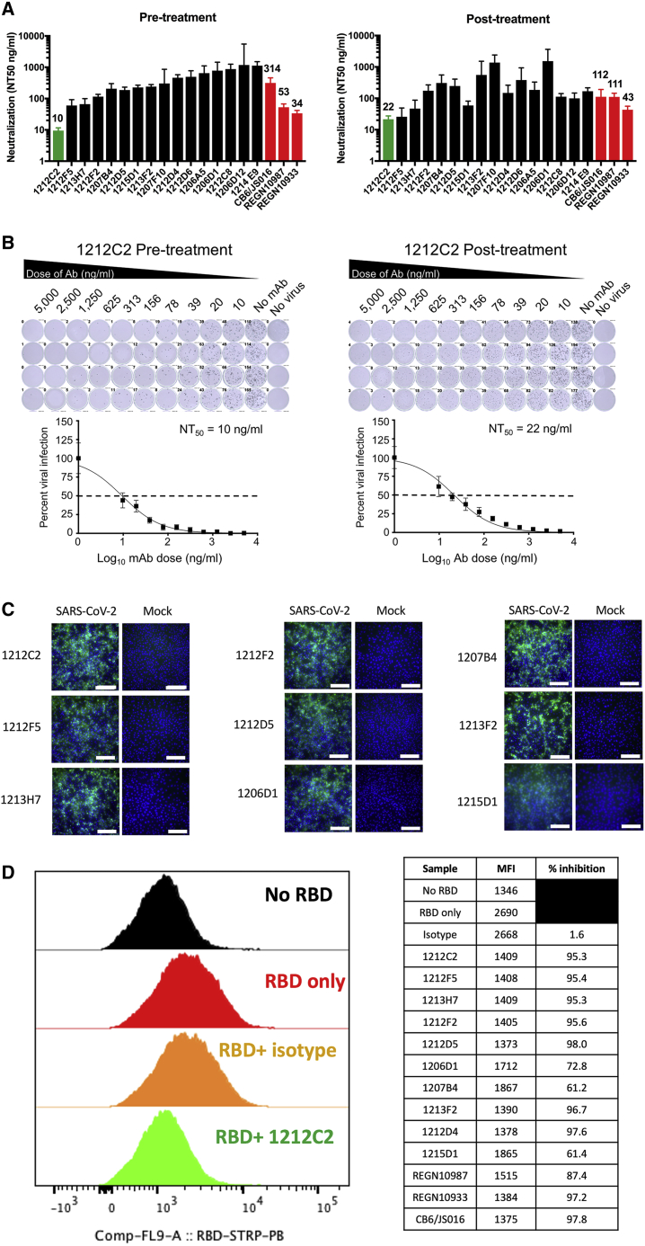
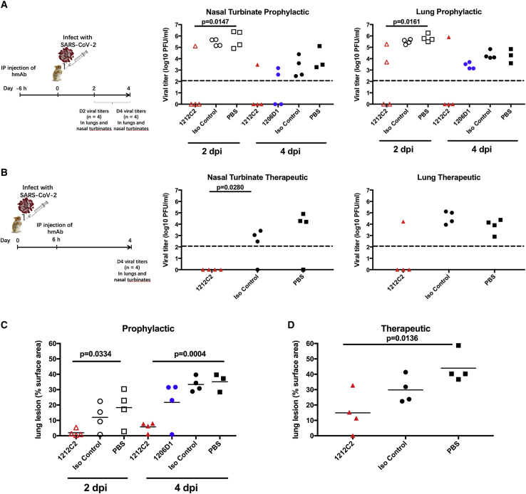
SARS-CoV-2 infection results in viral burden in the respiratory tract, enabling transmission and leading to substantial lung pathology. The 1212C2 fully human monoclonal antibody was derived from an IgM memory B cell of a COVID-19 patient, has high affinity for the Spike protein receptor binding domain, neutralizes SARS-CoV-2, and exhibits in vivo prophylactic and therapeutic activity in hamsters when delivered intraperitoneally, reducing upper and lower respiratory viral burden and lung pathology. Inhalation of nebulized 1212C2 at levels as low as 0.6 mg/kg, corresponding to 0.03 mg/kg lung-deposited dose, reduced the viral burden below the detection limit and mitigated lung pathology. The therapeutic efficacy of an exceedingly low dose of inhaled 1212C2 supports the rationale for local lung delivery for dose-sparing benefits, as compared to the conventional parenteral route of administration. These results suggest that the clinical development of 1212C2 formulated and delivered via inhalation for the treatment of SARS-CoV-2 infection should be considered.

Keywords: B cell; COVID-19; IgM; RBD; SARS-CoV-2; Spike; hamsters; inhalation; monoclonal antibody; neutralizing.


Conflict of interest statement

M.S.P., J.-G.P., A.D., F.S.O., M.B., S.S., N.B.E., P.A.G., M.R.W., L.M.-S., and J.J.K. are co-inventors on a patent that includes claims related to the hmAbs described. A.L., J.W., P.L., D.S., and V.L.T. are employees of Aridis Pharmaceuticals.

Figures

None
Graphical abstract
Figure 1
Figure 1
Isolation of SARS-CoV-2 RBD-specific human monoclonal antibodies (hmAbs) (A) Representative gating strategy for RBD+ B cell isolation. Initial plots are gated on live CD3CD4 CD19+ annexin V-CD27+ B cells. (B) hmAbs were tested at 10 and 1 μg/mL in duplicate by ELISA for binding to indicated protein; area under the curve (AUC) is indicated. (C) hmAbs were tested at 10 μg/mL in duplicate for binding to indicated protein by ELISA in the presence or absence of 8 M urea treatment, mean and standard error presented. (D) Binding to SARS-CoV-2 RBD was determined by surface plasmon resonance (SPR), mean and standard deviation presented.
Figure 2
Figure 2
Neutralization of SARS-CoV-2 infection and RBD binding to ACE2 (A) Indicated hmAbs were incubated with live SARS-CoV-2 (100 PFU/well) 1 h before (pre-treatment) or 1 h after (post-treatment) addition to Vero E6 cells. hmAbs were tested in quadruplicate cultures and NT50 and upper 95% confidence interval (CI) indicated. (B) Representative titration curve of 1212C2 hmAb presented, mean and standard error presented. (C) Binding of indicated hmAb to SARS-CoV-2 or mock-infected Vero E6 cells measured by immunofluorescence; scale bar, 100 μm. (D) Indicated hmAb was incubated as single replicate with recombinant biotinylated RBD protein before incubation with HEK293-ACE2 cells measured by flow cytometry. Plot gated on 7-aminoactinomycin (7AAD)-ACE2+ cells.
Figure 3
Figure 3
Phylogenic analysis of 1212C2 hmAb lineage Lineage members defined as same heavy-chain V and J gene usage, HCDR3 length, and ≥85% HCDR3 similarity. Germline sequence is represented by the green diamond, the 1212C2 hmAb sequence is represented by the red circle, and the 1212F5 hmAb sequenced is represented by the orange circle. The size of the symbols is proportional to the number of identical sequences obtained of an individual lineage member (N = 2–50), with the exception of the germline and 1212C2 and 1212F5 hmAb sequences. The relationship between lineage members was determined by amino acid sequence similarity.
Figure 4
Figure 4
SPR epitope mapping (A) Representative sensorgram from the SPR competition assays used to subset hmAbs into distinct RBD binding epitopes. For each assay, a series of hmAbs were sequentially injected over immobilized SARS-CoV-2 RBD. In this example, 1212C2 was injected first, followed by a second injection of 1212C2, 2 injections of 1206D1, and the last injection was CR3022. (B) Summary of all epitope mapping data, in which each block (first experiment from A in the red box) with a bold hmAb at the top represents a different experiment (10 experiments total). The bold hmAb is the “first” hmAb injected. The percentage of binding (100 = 100% binding and 0 = 0% binding) of subsequent hmAbs was recorded. mAbs were considered to have a different epitope (denoted by a distinct color) if they exhibited binding levels >30% in the presence of other mAbs. Thus, in the first experiment, CR3022 is defined as a new epitope (cyan), distinct from 1212C2. (C) Schematic diagram of NmAb RBD epitopes defined in the mapping experiment. Five major epitopes (A–E) were identified, where the E epitope overlaps with control mAb CR3022 (cyan, epitope F). Four of the 5 epitopes (A–D) are located within the ACE2 binding site (purple), and all of the NmAbs are blocked by the 1212C2 epitope A (yellow). NmAbs with epitopes similar to B (orange) and C (green) are defined as B’ (light orange) and C’ (light green), respectively. The 1212C2 epitope A (yellow) blocks the binding of all NmAbs, with the exception of 1215B11, which occupies epitope E. Epitopes B and C are also blocked by epitope A NmAbs, but exhibit limited competition with each other.
Figure 5
Figure 5
Prophylactic and therapeutic activity of 1212C2 hmAb in SARS-CoV-2-infected hamsters (A and B) Golden Syrian hamsters were (A) prophylactically treated i.p. with 10 mg/kg indicated hmAb or PBS 6 h before intranasal (i.n.) challenge with 2 × 105 PFU SARS-CoV-2 or (B) therapeutically treated i.p. with 25 mg/kg indicated hmAb or PBS 6 h following i.n. challenge with SARS-CoV-2. Virus present in nasal turbinates and lungs was determined by plaque assay. Dotted line indicates limit of detection. (C and D) Distribution of pathologic lesion, including consolidation, congestion, and pneumonic lesions, was measured using ImageJ and represented as the percentage of the total lung surface area in prophylactically treated (C) and therapeutically treated (D) animals. Each symbol represents an individual animal. p values determined by 2-tailed t test.
Figure 6
Figure 6
Therapeutic activity of inhaled 1212C2 hmAb in SARS-CoV-2-infected hamsters (A and B) Golden Syrian hamsters were infected i.n. with 2 × 105 PFU SARS-CoV-2 and 12 h later treated with indicated hmAb by i.p. or IH administration. Virus present in nasal turbinates and lungs was determined by plaque assay at 2 dpi (A) and 4 dpi (B). Dotted line indicates limit of detection. (C) Distribution of pathologic lesion were measured using ImageJ and represented as the percentage of the total lung surface area. ∗p < 0.0055 and ∗∗p < 0.00055 compared to PBS i.p.-treated group determined by 2-tailed t test.

References

    1. Cai Q., Chen F., Wang T., Luo F., Liu X., Wu Q., He Q., Wang Z., Liu Y., Liu L. Obesity and COVID-19 Severity in a Designated Hospital in Shenzhen, China. Diabetes Care. 2020;43:1392–1398. - PubMed
    1. Zhou F., Yu T., Du R., Fan G., Liu Y., Liu Z., Xiang J., Wang Y., Song B., Gu X. Clinical course and risk factors for mortality of adult inpatients with COVID-19 in Wuhan, China: a retrospective cohort study. Lancet. 2020;395:1054–1062. - PMC - PubMed
    1. CDC COVID-19 Response Team Preliminary Estimates of the Prevalence of Selected Underlying Health Conditions Among Patients with Coronavirus Disease 2019 - United States, February 12-March 28, 2020. MMWR Morb. Mortal. Wkly. Rep. 2020;69:382–386. - PMC - PubMed
    1. Wu C., Chen X., Cai Y., Xia J., Zhou X., Xu S., Huang H., Zhang L., Zhou X., Du C. Risk Factors Associated With Acute Respiratory Distress Syndrome and Death in Patients With Coronavirus Disease 2019 Pneumonia in Wuhan, China. JAMA Intern. Med. 2020;180:934–943. - PMC - PubMed
    1. Huang C., Wang Y., Li X., Ren L., Zhao J., Hu Y., Zhang L., Fan G., Xu J., Gu X. Clinical features of patients infected with 2019 novel coronavirus in Wuhan, China. Lancet. 2020;395:497–506. - PMC - PubMed

Publication types

MeSH terms