. 2021 May;23(5):309.
doi: 10.3892/mmr.2021.11948. Epub 2021 Mar 2.

Downregulation of lncRNA RPLP0P2 inhibits cell proliferation, invasion and migration, and promotes apoptosis in colorectal cancer

Affiliations

Downregulation of lncRNA RPLP0P2 inhibits cell proliferation, invasion and migration, and promotes apoptosis in colorectal cancer

Hang Yuan et al. Mol Med Rep. 2021 May.

Abstract

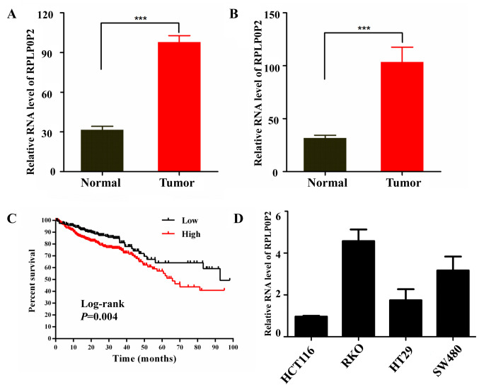
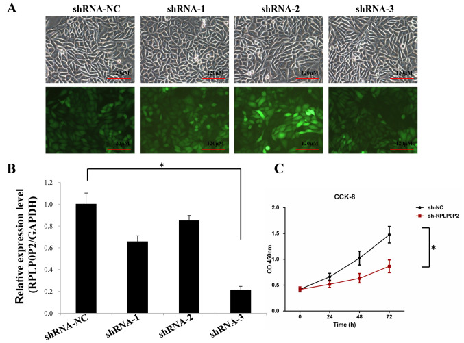
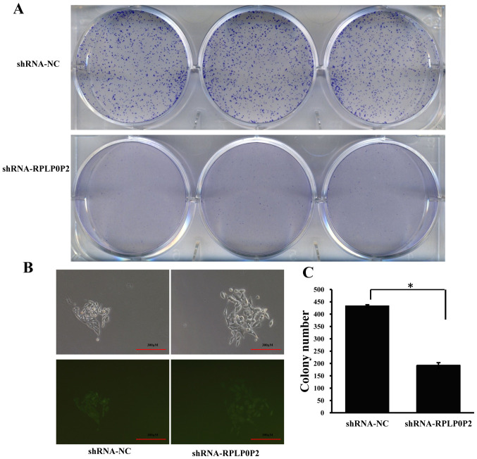
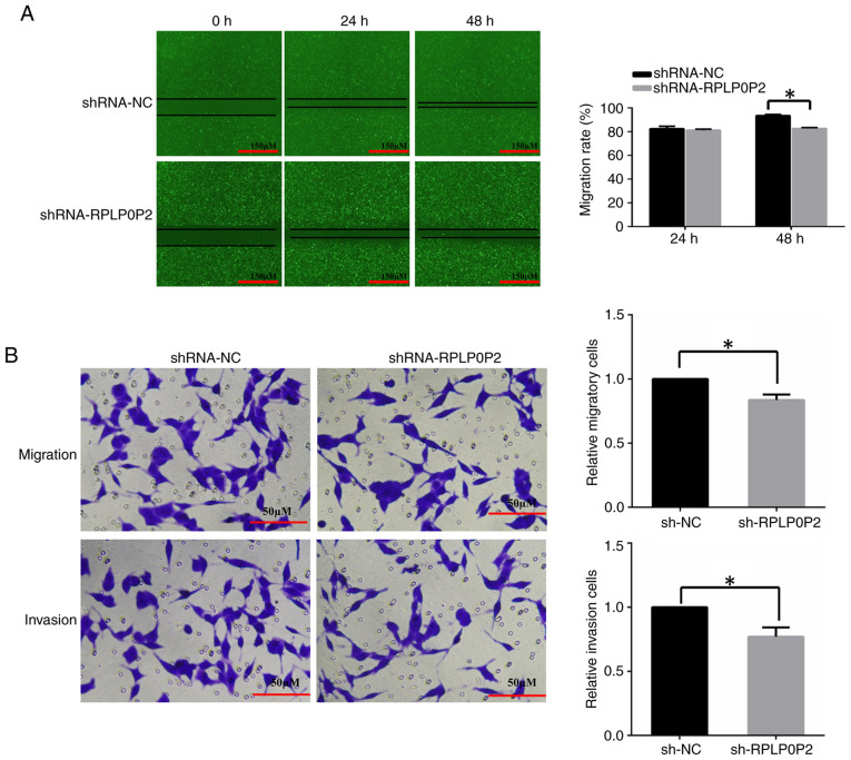
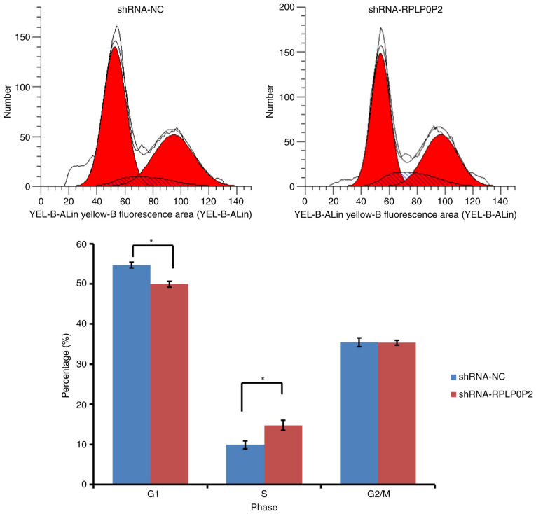
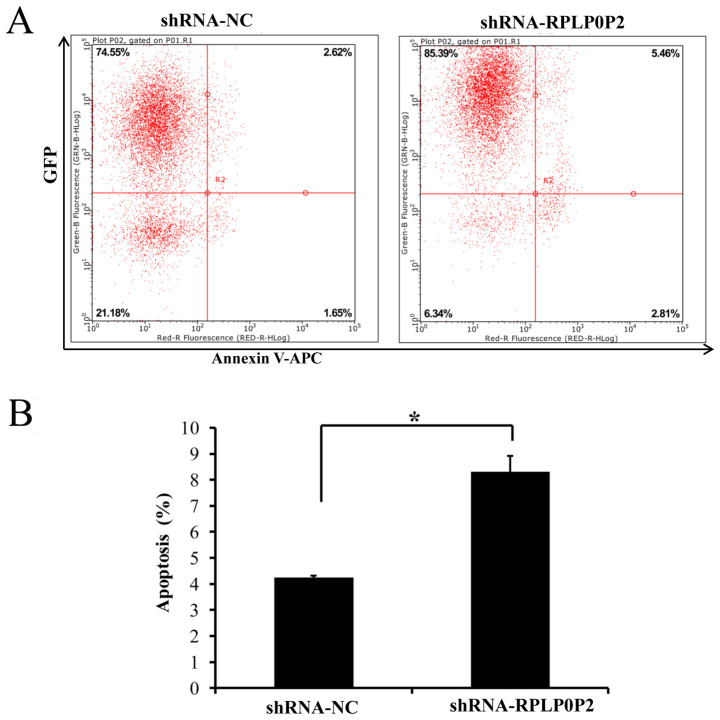
Recent studies have revealed that long noncoding RNAs (lncRNAs) are closely associated with colorectal cancer (CRC); however, the role of the lncRNA RPLP0P2 in CRC remains largely unknown. In the present study, RNA expression profiles of CRC were collected from The Cancer Genome Atlas database and the prognosis of CRC with respect to RPLP0P2 was assessed. Subsequently, RPLP0P2 expression was knocked down in the human CRC cell line RKO using a short hairpin RNA (shRNA) lentivirus, and the biological behaviors of the cells, such as proliferation, migration, cell cycle progression and apoptosis, were examined. The results demonstrated that the expression levels of RPLP0P2 were higher in CRC tissue compared with those in normal tissue, and RPLP0P2 was associated with prognosis. RPLP0P2 knockdown significantly decreased cell colony formation, migration and invasion, and arrested CRC cells in the S phase to G2/M phase transition. Furthermore, apoptosis was significantly increased in CRC cells infected with the RPLP0P2 shRNA lentivirus compared with in the control group. In conclusion, RPLP0P2 may promote proliferation, invasion and migration, and inhibit apoptosis of CRC cells, suggesting that RPLP0P2 may function as an oncogene in CRC.

Keywords: RPLP0P2; colorectal cancer; proliferation; apoptosis.


Conflict of interest statement

The authors declare that they have no competing interests.

Figures

Figure 1.
Figure 1.
Long non-coding RNA RPLP0P2 levels are elevated in CRC tissues. RPLP0P2 expression data in CRC tissues were obtained from The Cancer Genome Atlas database. (A) RPLP0P2 expression levels were higher in CRC tissues compared with in normal tissues (analysis of 51 cases of normal tissue and 647 cases of CRC tissue; test of two independent samples (Mann Whitney U test). (B) RPLP0P2 expression levels in 50 cases of matched CRC and normal tissue samples (test of paired samples, Wilcoxon signed-rank test). (C) High RPLP0P2 levels are associated with poor prognosis in patients with CRC. (D) Relative expression levels of RPLP0P2 in CRC cell lines. ***P<0.001. CRC, colorectal cancer.
Figure 2.
Figure 2.
Knockdown efficiency of RPLP0P2 with shRNA lentivirus in RKO cells. (A) Transfection efficiency of the RPLP0P2 shRNA lentivirus in RKO cells was ~80%. Upper rows, bright-field images of lentivirus infection; lower rows, fluorescent images of lentivirus infection. (B) Relative expression levels of RPLP0P2 were detected and it was revealed that the knockdown efficiency of shRNA-3 was the best. (C) CCK-8 cell proliferation assay revealed that shRNA-RPLP0P2 knockdown significantly decreased proliferation of RKO cells. *P<0.05. shRNA, short hairpin RNA; NC, negative control; CCK-8, Cell Counting Kit-8; OD, optical density.
Figure 3.
Figure 3.
RPLP0P2 knockdown inhibits cell colony formation. (A) Colony formation of RKO cells, which were stained with crystal violet. (B) Colony formation observed microscopically. Upper rows, bright field; lower rows: fluorescent view. (C) Number of stained colonies in RKO cells following infection with the RPLP0P2 shRNA lentivirus. Data are presented as the mean ± SD. *P<0.05. shRNA, short hairpin RNA; NC, negative control.
Figure 4.
Figure 4.
RPLP0P2 knockdown inhibits cell migration and invasion. (A) Wound area of the shRNA RPLP0P2 group was larger than in the control group after 48 h. Representative images and statistical analysis of the wound-healing assay are shown. Magnification, ×200. (B) Migration and invasion were reduced in the shRNA RPLP0P2 group compared with in the control group. Representative images of Transwell assay are shown. Magnification, ×200. *P<0.05. shRNA/sh, short hairpin RNA; NC, negative control.
Figure 5.
Figure 5.
Flow cytometric analysis of cell cycle progression. RPLP0P2 shRNA lentivirus enhanced the proportion of RKO cells in S phase and reduced the proportion of cells in G1 phase. *P<0.05. shRNA, short hairpin RNA; NC, negative control.
Figure 6.
Figure 6.
Analysis of apoptosis in lentivirus-infected RKO cells. (A) Apoptosis of RKO cells detected by flow cytometry following infection with the RPLP0P2 shRNA lentivirus. Apoptotic cells are shown in the top right and bottom right quadrants. Y-axis represents the lentivirus carrying enhanced green fluorescent protein transfected cells. X-axis represents APC-stained cells. (B) Apoptotic rate of RKO cells after infection with the RPLP0P2 shRNA lentivirus. *P<0.05. shRNA, short hairpin RNA; NC, negative control.

Similar articles

Cited by

References

    1. McGuire S. World cancer report 2014. Geneva, Switzerland: World health organization, international agency for research on cancer, WHO Press, 2015. Adv Nutr. 2016;7:418–419. doi: 10.3945/an.116.012211. - DOI - PMC - PubMed
    1. Gu MJ, Huang QC, Bao CZ, Li YJ, Li XQ, Ye D, Ye ZH, Chen K, Wang JB. Attributable causes of colorectal cancer in China. BMC Cancer. 2018;18:38. doi: 10.1186/s12885-017-3968-z. - DOI - PMC - PubMed
    1. Siegel RL, Miller KD, Fedewa SA, Ahnen DJ, Meester RGS, Barzi A, Jemal A. Colorectal cancer statistics, 2017. CA Cancer J Clin. 2017;67:177–193. doi: 10.3322/caac.21395. - DOI - PubMed
    1. Sana J, Faltejskova P, Svoboda M, Slaby O. Novel classes of non-coding RNAs and cancer. J Transl Med. 2012;10:103. doi: 10.1186/1479-5876-10-103. - DOI - PMC - PubMed
    1. Gong Z, Zhang S, Zhang W, Huang H, Li Q, Deng H, Ma J, Zhou M, Xiang J, Wu M, et al. Long non-coding RNAs in cancer. Sci China Life Sci. 2012;55:1120–1124. doi: 10.1007/s11427-012-4413-9. - DOI - PubMed

Substances