. 2021 Apr;32(4):958-969.
doi: 10.1111/jce.14980. Epub 2021 Mar 10.

Safety and chronic lesion characterization of pulsed field ablation in a Porcine model

Affiliations

Safety and chronic lesion characterization of pulsed field ablation in a Porcine model

Mark T Stewart et al. J Cardiovasc Electrophysiol. 2021 Apr.

Abstract

Background: Pulsed field ablation (PFA) has been identified as an alternative to thermal-based ablation systems for treatment of atrial fibrillation patients. The objective of this Good Laboratory Practice (GLP) study was to characterize the chronic effects and safety of overlapping lesions created by a PFA system at intracardiac locations in a porcine model.

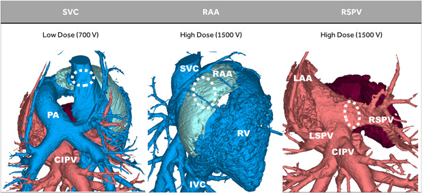
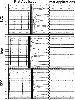
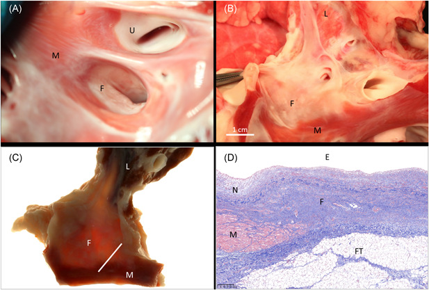
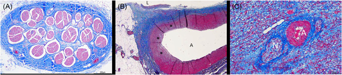
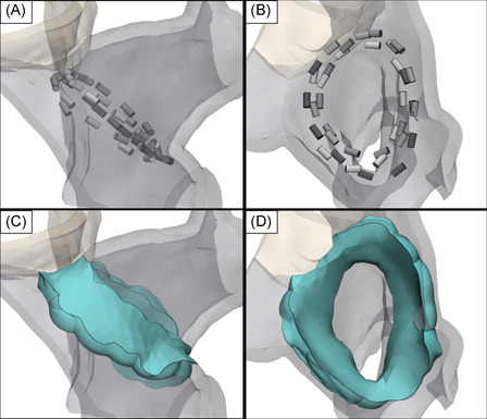
Methods: A circular catheter with nine gold electrodes was used for overlapping low- or high-dose PFA deliveries in the superior vena cava (SVC), right atrial appendage (RAA), and right superior pulmonary vein (RSPV) in six pigs. Electrical isolation was evaluated acutely and chronic lesions were assessed via necropsy and histopathology after 4-week survival. Acute and chronic safety data were recorded peri- and post-procedurally.

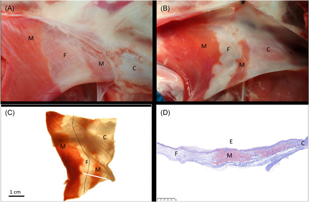
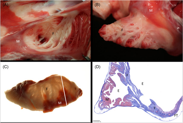
Results: No animal experienced ventricular arrhythmia during PFA delivery, and there was no evidence of periprocedural PFA-related adverse events. Lesions created in all anatomies resulted in electrical isolation postprocedure. Lesions were circumferential, contiguous, and transmural, with all converting into consistent lines of chronic replacement fibrosis, regardless of trabeculated or smooth endocardial surface structure. Ablations were non-thermally generated with only minimal post-delivery temperature rises recorded at the electrodes. There was no evidence of extracardiac damage, stenosis, aneurysms, endocardial disruption, or thrombus.

Conclusion: PFA deliveries to the SVC, RAA, and RSPV resulted in complete circumferential replacement fibrosis at 4-week postablation with an excellent chronic myocardial and collateral tissue safety profile. This GLP study evaluated the safety and efficacy of a dosage range in preparation for a clinical trial and characterized the non-thermal nature of PFA.

Keywords: atrial fibrillation; catheter ablation; electroporation; pulsed electric field modeling; pulsed field ablation.


Conflict of interest statement

David Haines, Atul Verma, Damijan Miklavčič, and Bor Kos receive research and consultation funds from Medtronic, Inc. Nicole Kirchhof, Noah Barka, Lars Mattison, Matthew Martien, Birce Onal, Brian Howard, and Mark Stewart are employees of Medtronic, Inc.

Figures

Figure 1
Figure 1
Porcine model summary. In six pigs, a low (±700 V) dose of pulsed field ablation (PFA) was delivered to the superior vena cava (SVC) (muscle sleeve portion), and a high (±1500 V) dose of PFA was delivered to the right atrial appendage (RAA), and right superior pulmonary vein (RSPV). The common inferior pulmonary vein (CIPV) was not ablated
Figure 2
Figure 2
Acute Isolation of the SVC, RAA, and RSPV. Immediately after the first pulse train delivery, electrograms for each of the targeted sites were reduced and after all four pulse trains were delivered, only far‐field signals remained. Complete electrical isolation of all structures was confirmed by entrance block. Entrance and exit block were confirmed for the SVC. RAA, right atrial appendage; RSPV, right superior pulmonary vein; SVC, superior vena cava
Figure 3
Figure 3
Ablation of the SVC. In‐situ necropsy finding, enhanced by triphenyl tetrazolium chloride (TTC) staining, 4 weeks after low‐dose PFA treatment of the SVC before (A) and after (B) opening the lumen. A continuous ablation line (dotted line) is readily visible under backlight illumination of the same specimen after formalin fixation (C). Histology confirms transmural replacement fibrosis and shows abrupt transition to normal, nontreated muscle via Masson's trichrome staining. White bar in (C) indicates slide origin for (D). C, collagenous portion of the SVC; E, endothelial side; F, fibrosis after ablation; M, muscle (untreated); PFA, pulsed field ablation; SVC, superior vena cava
Figure 4
Figure 4
Ablation of the RAA. In‐situ necropsy finding, enhanced by TTC staining, 4 weeks after high‐dose PFA treatment of the RAA when looking into the appendage (A) and after eversion of the appendage to display the endocardium (B). Widespread atrial replacement fibrosis is readily visible under backlight illumination of the same specimen after formalin fixation (C). Histology via Masson's trichrome staining confirms transmural replacement fibrosis in atrial wall segments that were originally made of irregular trabeculation/pectination. White bar in (C) indicates slide origin for (D). E, endothelial side; F, fibrosis after ablation; FT, fat tissue on epicardium (normal); M, muscle (untreated); PFA, pulsed field ablation; RAA, right atrial appendage; TTC, triphenyl tetrazolium chloride
Figure 5
Figure 5
Ablation of the RSPV. In‐situ necropsy finding, enhanced by TTC staining, 4 weeks after high‐dose PFA treatment of the RSPV when looking into its orifice (A) and after longitudinal opening towards the lung (B). There is good evidence of circumferential replacement fibrosis when the same formalin‐fixed specimen is examined under backlight illumination (C). Histology via Masson's trichrome staining confirms transmural replacement fibrosis that is covered by neointima and has normal adjacent myocardium (D). White bar in (C) indicates slide origin for (D). E, endothelial side; F, fibrosis after ablation; FT, fat tissue at cardiac base (normal); L, lung; M, muscle (untreated); N, neointima; PFA, pulsed field ablation; RSPV, right superior pulmonary vein; TTC, triphenyl tetrazolium chloride; U, untreated pulmonary vein orifice
Figure 6
Figure 6
(A) Collateral damage profile. Photomicrograph of the right phrenic nerve that illustrates no pathological changes 4 weeks after PFA treatment when low‐dose PFA was delivered in the SVC. (B) Histology of a pulmonary artery branch previously juxtaposed to a RSPV ablation and now showing arterial wall remodeling that consisted of mild fibrosis and loss of media myocytes. Photomicrograph from a right atrial appendage with perineural fibrosis and normal appearing artery (C). A, artery; L, lumen; N, nerve; PFA, pulsed field ablation; RSPV, right superior pulmonary vein; SVC, superior vena cava. *Fibrosis and myocyte loss
Figure 7
Figure 7
Three‐dimensional rendering of the RSPV constructed using contrast enhanced CT images from a single pig. 4 placements of the circular electrode array were positioned at the ostium of the RSPV with the shape of the catheter conforming to the inner surface of the ostium (A and B). Using a conservative field threshold of 550 V/cm and uniform 3 mm tissue thickness, the field isosurfaces predict a contiguous and transmural lesion volume (green) with lesion widths ranging 3–12 mm on the outer surface of the ostium and 6.5–15 mm on the inner surface (C and D). CT, computed tomography; RSPV, right superior pulmonary vein

Comment in

References

    1. Chugh SS, Havmoeller R, Narayanan K, et al. Worldwide epidemiology of atrial fibrillation: a global burden of disease 2010 study. Circulation. 2014;129:837‐847. 10.1161/circulationaha.113.005119 - DOI - PMC - PubMed
    1. Morillo CA, Banerjee A, Perel P, Wood D, Jouven X. Atrial fibrillation: the current epidemic. J Geriatr Cardiol. 2017;14(3):195‐203. 10.11909/j.issn.1671-5411.2017.03.011 - DOI - PMC - PubMed
    1. Calkins H, Hindricks G, Cappato R, et al. HRS/EHRA/ECAS/APHRS/SOLAECE expert consensus statement on catheter and surgical ablation of atrial Fibrillation. Heart Rhythm. 2017;14:e275‐e444. 10.10106/j.hrthm.2017.05.012 - DOI - PMC - PubMed
    1. Haegeli LM, Calkins H. Catheter ablation of atrial fibrillation: an update. Eur Heart J. 2014;35(36):2454‐2459. 10.1093/eurheartj/ehu291 - DOI - PubMed
    1. Wilber DJ, Pappone C, Neuzil P, et al. Comparison of antiarrhythmic drug therapy and radiofrequency catheter ablation in patients with paroxysmal atrial fibrillation: a randomized controlled trial. JAMA. 2010;303(4):333‐340. 10.1001/jama.2009.2029 - DOI - PubMed

Publication types