Review
. 2021 Apr;25(8):3680-3698.
doi: 10.1111/jcmm.16397. Epub 2021 Mar 2.

Gynaecological cancers and their cell lines

Affiliations
Review

Gynaecological cancers and their cell lines

Kristijan Skok et al. J Cell Mol Med. 2021 Apr.

Abstract

Cell lines are widely used for various research purposes including cancer and drug research. Recently, there have been studies that pointed to discrepancies in the literature and usage of cell lines. That is why we have prepared a comprehensive overview of the most common gynaecological cancer cell lines, their literature, a list of currently available cell lines, and new findings compared with the original studies. A literature review was conducted via MEDLINE, PubMed and ScienceDirect for reviews in the last 5 years to identify research and other studies related to gynaecological cancer cell lines. We present an overview of the current literature with reference to the original studies and pointed to certain inconsistencies in the literature. The adherence to culturing rulesets and the international guidelines helps in minimizing replication failure between institutions. Evidence from the latest research suggests that despite certain drawbacks, variations of cancer cell lines can also be useful in regard to a more diverse genomic landscape.

Keywords: breast neoplasm; cell line; cervix cancer; endometrial neoplasms; gynaecology; in vitro techniques; pathology; tumour cell line.


Conflict of interest statement

The authors declare no conflict of interest. The funders had no role in the design of the study; in the collection, analyses or interpretation of data; in the writing of the manuscript; or in the decision to publish the results.

Figures

FIGURE 1
FIGURE 1
Potential CCL applications in research
FIGURE 2
FIGURE 2
PRISMA diagram of the conducted search inquiries
FIGURE 3
FIGURE 3
HeLa cell line. (A) Electron micrograph of an apoptotic HeLa cell (source: National Institutes of Health—NIH; https://imagebank.nih.gov/details.cfm?imageid=1463); (B) Immunofluorescence image of HeLa cells grown in tissue culture and stained with antibody to actin in green, vimentin in red and DNA in blue (source: GerryShaw—wikimedia.commons; https://commons.wikimedia.org/wiki/File:HeLa_cells_stained_with_antibody_to_actin_(green)_,_vimentin_(red)_and_DNA_(blue).jpg); (C) Multiphoton fluorescence image of cultured HeLa cells with a fluorescent protein targeted to the Golgi apparatus (orange), microtubules (green) and counterstained for DNA (cyan) (source: NIH; https://commons.wikimedia.org/wiki/File:HeLa‐I.jpg); D) Immunofluorescence of HeLa cells showing microtubules in green, mitochondria in yellow, nucleoli in red and nuclear DNA in purple (source: GerryShaw—wikimedia.commons; https://commons.wikimedia.org/wiki/File:HeLa‐Tubulin‐HSP60‐Fibrillarin‐DNA.jpg). All material is published under the CC license or is in the public domain
FIGURE 4
FIGURE 4
A simplified geographical overview of the percentage of contaminated primary articles as a fraction of the total number of articles on cells per country (Top 5: Japan, Brazil, Taiwan, India, China). Adapted from the dataset from Horbach and Halffman 68 under the CC license
FIGURE 5
FIGURE 5
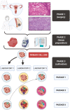
Cell line isolation and changes during cultivation. Phases 1 and 2 encompass the logistics of tissue retrieval and pre‐cultivation procedures (eg homogenization). Phase 3 shows the heterogeneity and susceptibility of CLs to mutate due to various changes. Laboratory 1 and laboratory 2 received commercially bought CLs. The CL from laboratory 1 mutated somewhere between passage 3 and 6. Laboratory 2 used from the beginning a different medium, which led to a variety of genotypical and phenotypical changes. Laboratory 3 borrowed a CL sample (already mutated) from laboratory 1. Due to careless handling, the CL got infected with mycoplasma. The addition of an antibiotic agent again led to a series of genotypical and phenotypical changes in the CL. Source: Histological images were used under the CC license from Wikimedia Commons (https://commons.wikimedia.org/wiki/File:Invasive_Ductal_Carcinoma_40x.jpg; https://commons.wikimedia.org/wiki/File:Endometrioid_endometrial_adenocarcinoma_very_high_mag.jpg). The figure itself was created with BioRender.com
FIGURE 6
FIGURE 6
Cell culturing protocol

References

    1. Galuschka C, Proynova R, Roth B, Augustin HG, Müller‐Decker K. Models in translational oncology: a public resource database for preclinical cancer research. Cancer Res. 2017;77(10):2557‐2563. 10.1158/0008-5472.CAN-16-3099 - DOI - PubMed
    1. Skok K, Maver U, Gradišnik L, Kozar N, Takač I, Arko D. Endometrial cancer and its cell lines. Mol Biol Rep. 2020;47(2):1399‐1411. 10.1007/s11033-019-05226-3 - DOI - PubMed
    1. Skok K, Gradišnik L, Čelešnik H, et al. Isolation and characterization of the first Slovenian human triple‐negative breast cancer cell line. Breast J. 2020;26(2):328‐330. 10.1111/tbj.13695 - DOI - PubMed
    1. Skok K, Maver U, Gradišnik L, Sobočan M, Takač I. Humane celične linije raka dojk. Slovenian Medical Journal. 2019;88 (9‐10):427–43. 10.6016/zdravvestn.2842 - DOI
    1. Lin CY, Erkek S, Tong Y, et al. Active medulloblastoma enhancers reveal subgroup‐specific cellular origins. Nature. 2016;530(7588):57‐62. 10.1038/nature16546 - DOI - PMC - PubMed

Publication types

LinkOut - more resources