Review
. 2021 Apr;299(1):36-48.
doi: 10.1148/radiol.2021201948. Epub 2021 Mar 2.

Contrast-enhanced Mammography: State of the Art

Affiliations
Review

Contrast-enhanced Mammography: State of the Art

Maxine S Jochelson et al. Radiology. 2021 Apr.

Abstract

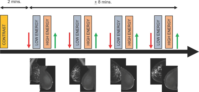
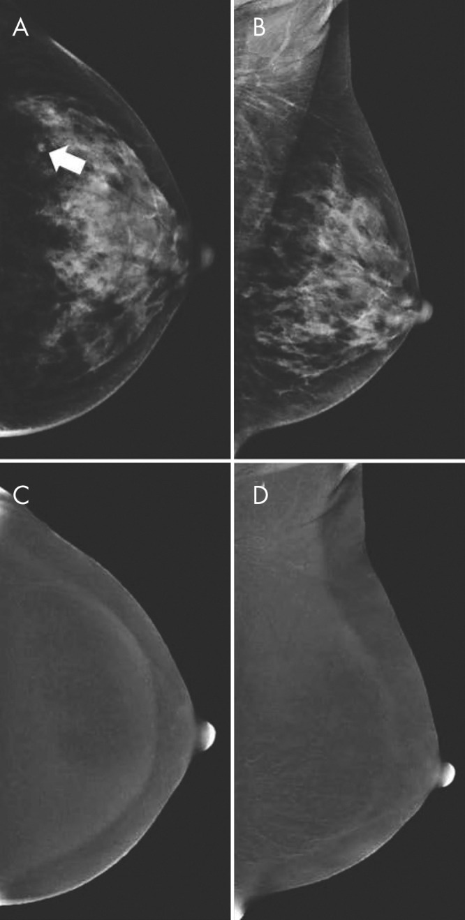
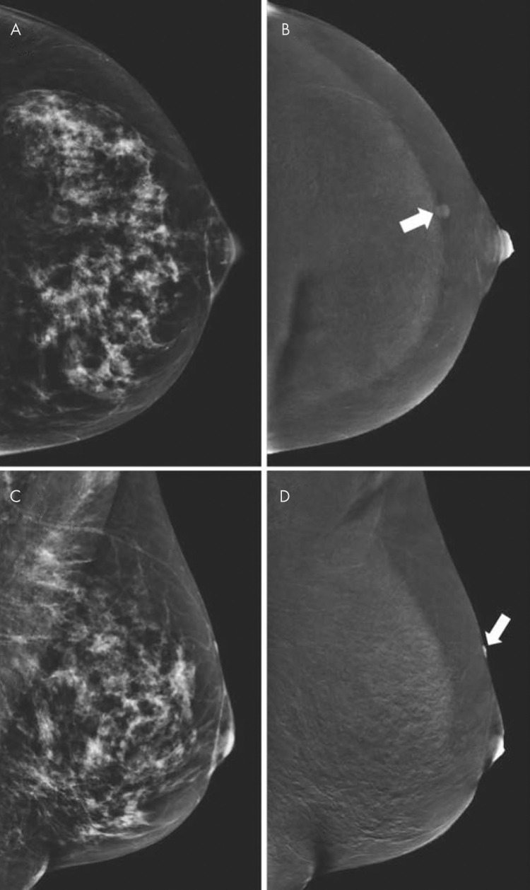
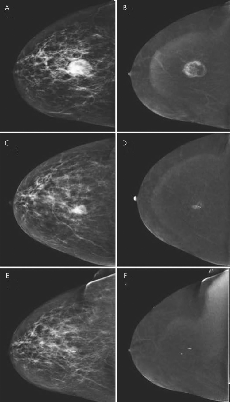
Contrast-enhanced mammography (CEM) has emerged as a viable alternative to contrast-enhanced breast MRI, and it may increase access to vascular imaging while reducing examination cost. Intravenous iodinated contrast materials are used in CEM to enhance the visualization of tumor neovascularity. After injection, imaging is performed with dual-energy digital mammography, which helps provide a low-energy image and a recombined or iodine image that depict enhancing lesions in the breast. CEM has been demonstrated to help improve accuracy compared with digital mammography and US in women with abnormal screening mammographic findings or symptoms of breast cancer. It has also been demonstrated to approach the accuracy of breast MRI in preoperative staging of patients with breast cancer and in monitoring response after neoadjuvant chemotherapy. There are early encouraging results from trials evaluating CEM in the screening of women who are at an increased risk of breast cancer. Although CEM is a promising tool, it slightly increases radiation dose and carries a small risk of adverse reactions to contrast materials. This review details the CEM technique, diagnostic and screening uses, and future applications, including artificial intelligence and radiomics.


Figures

None
Graphical abstract
Diagram of imaging protocol for contrast-enhanced mammography. Two minutes before image acquisition, iodine-based contrast material is injected. Next, at minimum, both breasts are imaged in craniocaudal and mediolateral oblique views. In each step, compression is applied (red arrow), followed by rapid acquisition of low- and high-energy images. These images are processed to generate low-energy and recombined images. After each exposure, compression is released (green arrow). Images are considered to be of diagnostic value if they are acquired within 10 minutes after contrast material administration. mins. = minutes.
Figure 1:
Diagram of imaging protocol for contrast-enhanced mammography. Two minutes before image acquisition, iodine-based contrast material is injected. Next, at minimum, both breasts are imaged in craniocaudal and mediolateral oblique views. In each step, compression is applied (red arrow), followed by rapid acquisition of low- and high-energy images. These images are processed to generate low-energy and recombined images. After each exposure, compression is released (green arrow). Images are considered to be of diagnostic value if they are acquired within 10 minutes after contrast material administration. mins. = minutes.
A, B, Contrast-enhanced mammographic (CEM) images in 52-year-old woman recalled from screening for new ill-defined mass (arrow in A) visible on, A, low-energy craniocaudal view but not visible on, B, low-energy mediolateral oblique view. C, D, Subsequent evaluation of recombined CEM images revealed no suspicious enhancement. Targeted US showed no lesions, but because of small lesion size, patient was categorized as having Breast Imaging Reporting and Data System category 3 lesion (follow-up after 6 months showed no breast cancer). Two subsequent rounds of screening (up to 4 years after primary evaluation) revealed no breast cancer.
Figure 2:
A, B, Contrast-enhanced mammographic (CEM) images in 52-year-old woman recalled from screening for new ill-defined mass (arrow in A) visible on, A, low-energy craniocaudal view but not visible on, B, low-energy mediolateral oblique view. C, D, Subsequent evaluation of recombined CEM images revealed no suspicious enhancement. Targeted US showed no lesions, but because of small lesion size, patient was categorized as having Breast Imaging Reporting and Data System category 3 lesion (follow-up after 6 months showed no breast cancer). Two subsequent rounds of screening (up to 4 years after primary evaluation) revealed no breast cancer.
Contrast-enhanced mammographic images of enhancing benign lesion in 50-year-old woman. A, Low-energy craniocaudal image. B, Contrast-enhanced recombined craniocaudal image. C, Low-energy mediolateral oblique image. D, Contrast-enhanced recombined mediolateral oblique image. Contrast-enhanced craniocaudal view of left breast demonstrates a small, well-defined enhancing mass (arrow in B). Mediolateral oblique view of same breast demonstrates that lesion is located on skin (arrow in D). Visual inspection revealed skin hemangioma.
Figure 3:
Contrast-enhanced mammographic images of enhancing benign lesion in 50-year-old woman. A, Low-energy craniocaudal image. B, Contrast-enhanced recombined craniocaudal image. C, Low-energy mediolateral oblique image. D, Contrast-enhanced recombined mediolateral oblique image. Contrast-enhanced craniocaudal view of left breast demonstrates a small, well-defined enhancing mass (arrow in B). Mediolateral oblique view of same breast demonstrates that lesion is located on skin (arrow in D). Visual inspection revealed skin hemangioma.
Images in 44-year-old woman presenting with palpable abnormality in left breast illustrate that cysts have different appearance at contrast-enhanced mammography (CEM) and contrast-enhanced breast MRI. A, Recombined CEM image of left breast in mediolateral oblique view. B, Corresponding contrast-enhanced fat-suppressed T1-weighted breast MRI scan reconstructed in sagittal view. On both images, simple cysts (arrowheads) can be identified as well-defined masses showing no or negative enhancement. Inflamed cysts (arrow) may show thickened and often slightly irregular wall, which enhances after contrast material administration.
Figure 4:
Images in 44-year-old woman presenting with palpable abnormality in left breast illustrate that cysts have different appearance at contrast-enhanced mammography (CEM) and contrast-enhanced breast MRI. A, Recombined CEM image of left breast in mediolateral oblique view. B, Corresponding contrast-enhanced fat-suppressed T1-weighted breast MRI scan reconstructed in sagittal view. On both images, simple cysts (arrowheads) can be identified as well-defined masses showing no or negative enhancement. Inflamed cysts (arrow) may show thickened and often slightly irregular wall, which enhances after contrast material administration.
Images obtained for disease extent assessment in 64-year-old woman recalled from screening for irregular mass in right breast. A, Low-energy image of right breast (craniocaudal view) shows irregular mass (arrow). B, No lesion is seen on low-energy image in contralateral breast. C, Image from contrast-enhanced mammography (CEM) (right craniocaudal view) shows enhancement of mass (arrow). D, CEM image shows contralateral irregular enhancing mass (arrow), which was not visible on low-energy image. E, F, Contrast-enhanced MRI scans of, E, right breast and, F, left breast show both lesions (arrow). Lesions were diagnosed as invasive breast cancer of no special type (estrogen receptor positive, progesterone receptor positive, and human epidermal growth factor receptor type 2 negative).
Figure 5:
Images obtained for disease extent assessment in 64-year-old woman recalled from screening for irregular mass in right breast. A, Low-energy image of right breast (craniocaudal view) shows irregular mass (arrow). B, No lesion is seen on low-energy image in contralateral breast. C, Image from contrast-enhanced mammography (CEM) (right craniocaudal view) shows enhancement of mass (arrow). D, CEM image shows contralateral irregular enhancing mass (arrow), which was not visible on low-energy image. E, F, Contrast-enhanced MRI scans of, E, right breast and, F, left breast show both lesions (arrow). Lesions were diagnosed as invasive breast cancer of no special type (estrogen receptor positive, progesterone receptor positive, and human epidermal growth factor receptor type 2 negative).
Images of response monitoring using contrast-enhanced mammography in a 54-year-old woman diagnosed with 3.0-cm invasive breast cancer of no special type (grade 3, estrogen receptor negative, progesterone receptor negative, and human epidermal growth factor receptor type 2 positive). A, Low-energy image in craniocaudal view shows cancer. B, Recombined image shows peripheral enhancement, which is suggestive of central necrosis. The patient underwent neoadjuvant systemic therapy. C, Low-energy, and, D, recombined craniocaudal views obtained during treatment show decrease in index tumor size (C) with decrease in enhancement (D). E, Low-energy and, F, recombined images obtained after completion of therapy. There was no residual mass on low-energy image. Resolution of enhancement on recombined image was consistent with complete radiologic response. Surgical specimen showed pathologic complete response without any residual ductal carcinoma in situ.
Figure 6:
Images of response monitoring using contrast-enhanced mammography in a 54-year-old woman diagnosed with 3.0-cm invasive breast cancer of no special type (grade 3, estrogen receptor negative, progesterone receptor negative, and human epidermal growth factor receptor type 2 positive). A, Low-energy image in craniocaudal view shows cancer. B, Recombined image shows peripheral enhancement, which is suggestive of central necrosis. The patient underwent neoadjuvant systemic therapy. C, Low-energy, and, D, recombined craniocaudal views obtained during treatment show decrease in index tumor size (C) with decrease in enhancement (D). E, Low-energy and, F, recombined images obtained after completion of therapy. There was no residual mass on low-energy image. Resolution of enhancement on recombined image was consistent with complete radiologic response. Surgical specimen showed pathologic complete response without any residual ductal carcinoma in situ.

References

    1. Patel BK, Lobbes MBI, Lewin J. Contrast Enhanced Spectral Mammography: A Review. Semin Ultrasound CT MR 2018;39(1):70–79. - PubMed
    1. Kuhl C. The current status of breast MR imaging. Part I. Choice of technique, image interpretation, diagnostic accuracy, and transfer to clinical practice. Radiology 2007;244(2):356–378. - PubMed
    1. Carney PA, Miglioretti DL, Yankaskas BC, et al. Individual and combined effects of age, breast density, and hormone replacement therapy use on the accuracy of screening mammography. Ann Intern Med 2003;138(3):168–175. - PubMed
    1. Weigel S, Heindel W, Heidrich J, Hense HW, Heidinger O. Digital mammography screening: sensitivity of the programme dependent on breast density. Eur Radiol 2017;27(7):2744–2751. - PubMed
    1. Tagliafico AS, Calabrese M, Mariscotti G, et al. Adjunct Screening With Tomosynthesis or Ultrasound in Women With Mammography-Negative Dense Breasts: Interim Report of a Prospective Comparative Trial. J Clin Oncol 2016;34(16):1882–1888. - PubMed

Publication types

LinkOut - more resources