Review
. 2022 Aug;42(6):1671-1692.
doi: 10.1007/s10571-021-01064-9. Epub 2021 Mar 2.

The Roles of Serotonin in Neuropsychiatric Disorders

Affiliations
Review

The Roles of Serotonin in Neuropsychiatric Disorders

Mahsa Pourhamzeh et al. Cell Mol Neurobiol. 2022 Aug.

Abstract

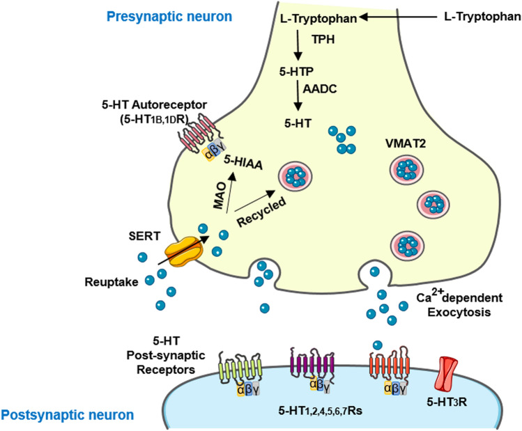
The serotonergic system extends throughout the central nervous system (CNS) and the gastrointestinal (GI) tract. In the CNS, serotonin (5-HT, 5-hydroxytryptamine) modulates a broad spectrum of functions, including mood, cognition, anxiety, learning, memory, reward processing, and sleep. These processes are mediated through 5-HT binding to 5-HT receptors (5-HTRs), are classified into seven distinct groups. Deficits in the serotonergic system can result in various pathological conditions, particularly depression, schizophrenia, mood disorders, and autism. In this review, we outlined the complexity of serotonergic modulation of physiologic and pathologic processes. Moreover, we provided experimental and clinical evidence of 5-HT's involvement in neuropsychiatric disorders and discussed the molecular mechanisms that underlie these illnesses and contribute to the new therapies.

Keywords: 5-HT; Mood Disorders; Nervous System Diseases; SSRI; Serotonin pathway.


Conflict of interest statement

The authors declare no conflict of interest regarding the publication of this paper.

Figures

Fig. 1
Fig. 1
5-HT Synthesis and Metabolism. The essential amino acid, tryptophan, is the sole precursor molecule involved with the synthesis of 5-HT. Tryptophan must first cross the BBB to enter the CNS prior to 5-HT synthesis. Once in the CNS, l-tryptophan is hydroxylated to 5-hydroxytryptophan (5-HTP) by the enzyme tryptophan hydroxylase type 2 (TPH2). This is followed by subsequent decarboxylation involving the enzyme l-aromatic acid decarboxylase (AADC) transforming 5-HTP into 5-HT. This fully synthesized 5-HT is then taken up into vesicles in the axon terminal via vesicular monoamine transporter isoform 2 (VMAT2). Following an action potential, 5-HT is released into the synapse. It can interact with both presynaptic and postsynaptic receptors. In addition, 5-HT can be metabolized by monoamine oxidase-A (MAO-A) and aldehyde dehydrogenase to the metabolite 5-hydroxyindoleacetic acid (5-HIAA). 5-HT synthesis is regulated by the tryptophan-degrading enzyme, indoleamine 2,3-dioxygenase, and the cofactor of tryptophan hydroxylase, tetrahydrobiopterin. All 5-HTRs are heteroreceptors and postsynaptically expressed on non-serotonergic neurons and autoreceptors located presynaptically on the serotonergic neurons. SERTs are localized on the axon terminal and soma of the serotonergic neurons. SERT undergoes conformational modifications and transfers one or more molecules in each cycle (El-Merahbi et al. ; Sahu et al. 2018)
Fig. 2
Fig. 2
Summary of proposed signaling mediated by 5-HT receptor subtypes. 5-HT1A, B, D, E, F, 5-HT2A,B,C, 5-HT4, 5-HT5A, B, 5-HT6, and 5-HT7 receptors are classified as G protein-coupled receptors (GPCRs), while 5-HT3A, B, C, D, E receptors are ligand-gated ion channels (LGIC). The serotonergic GPCRs are made of a common structure and contain seven transmembrane alpha-helices (7-TM), which are connected by three extracellular and three intracellular loop. Upon ligand binding, the intracellular loop and C-terminal tail interact with specific G protein families, including Gαs, Gαi/o, Gαq/11, and followed by the generation of second messengers. 5-HT1Rs couple to Gαi/o proteins, 5-HT2Rs couple to Gαq/11 proteins, and 5-HT4, 5-HT6 and 5-HT7 receptors couple to Gαs proteins. Although no primary mechanistic pathway has been recognized for 5-HT5Rs, there are reports suggesting that it couples to Gαi/o. 5-HT3Rs are made of five monomers (A to E) and form a tube-like channel. These GPCRs work through a variety of intracellular functional proteins, including adenylyl cyclase (AC), Phospholipase-C (PLC), voltage-gated N-type Ca2+ channels, and hyperpolarizing K+ channels. 5-HT1Rs inhibit neuronal activity by suppressing the activity of AC, and decreasing the cAMP level, and activate MAPK pathway. Coupling through 5-HT2Rs induces PLC activity which hydrolyze membrane phospholipids to release second messengers, such as inositol 1,4,5-triphosphate (IP3) and diacylglycerols (DAGs). It also stimulates protein kinase C (PKC), MAPK pathway, and hyperpolarizes K+ channels. The activation of 5-HT5ARs activate the formation of cyclic ADP ribose (cADPR), and in turn elicit Ca2+ release. However, 5-HT4, 5-HT6, and 5-HT7 receptors stimulate AC, resulting in increased cAMP level, activated protein kinase A (PKA), and the regulation of several signaling molecules, such as CREB. The MAPK pathway concludes with the activation of Erk1 and Erk2 proteins which in turn enter the nucleus and cause phosphorylation-mediated activation of transcription factors, such as Elk1, and CREB. When 5-HT binds to 5-HT3Rs, a gate opens up and becomes permeable to Na+, K+, and Ca2+ ions. Ca2+ decreases 5-HT-induced Na+ currents in a concentration-dependent manner without affecting the internal alteration of the current/voltage relation (Adayev et al. ; Filip and Bader ; Engel et al. ; Locher et al. 2017)
Fig. 3
Fig. 3
Serotonergic Pathways. In addition to the dorsal and median raphe nuclear complex, many serotonergic neurons are found in the reticular region of the lower brain stem, which is subdivided into caudal and rostral groups. Serotonergic neurons of the rostral system (caudal linear, dorsal raphe, and median raphe nuclei) mostly terminate in the dorsal and medial raphe nuclei, which in turn innervate much of the rest of the CNS by diffuse projections. Serotonergic neurons of the caudal group (raphe magnus, raphe obscurus, raphe pallidus nuclei) descend into the brain stem and the spinal cord along the dorsal horn, ventral horn motor nuclei and thoracic cord intermediolateral column and are mostly involved in sensory, motor and autonomic functioning. The central gray matter and the ependyma of the central canal also receives 5-HT input in a pattern of 5-HT nerve plexus. Serotonergic signaling regulates impulsivity and behavioral adaptation through actions within the medial prefrontal cortex (mPFC). Serotonergic innervation is present throughout the NAc and involved in social behaviors. Moreover, serotonergic fibers innervate the amygdala and enhance aversive memory acquisition. Signaling of 5-HT in the periaqueductal gray (PAG) is involved in behavioral inhibition and prevent panic-like behaviors. Additionally, serotonergic innervation is present throughout the bed nucleus of the stria terminalis (BNST) and could promote or inhibit approach and avoidance behaviors. The expression of the fear- and anxiety-related defensive behavioral responses of the habenula (Hb) is also regulated by serotonergic projections. The presence of 5-HT has been observed in the pons, medulla, septal area and specific areas of the thalamus, hypothalamus, substantia nigra (SN) and locus coeruleus (LC). The hippocampus (Hippo), the suprachiasmatic nucleus, the olfactory bulb and the medial septum nucleus receive 5-HT innervation from the MRN. Likewise, serotonergic fibers are one of the afferent fibers innervating and affecting the activity of cerebellar circuitry (Dölen ; Jorgensen ; Puig and Gulledge ; Linley et al. ; Saitow et al. 2012)
Fig. 4
Fig. 4
A summary of the major nervous system diseases associated with dysfunctions in serotonergic receptors

References

    1. Abela AR, Browne CJ, Sargin D, Prevot TD, Ji XD, Li Z, Lambe EK, Fletcher PJ (2020) Median raphe serotonin neurons promote anxiety-like behavior via inputs to the dorsal hippocampus. Neuropharmacology 15(168):107985. 10.1016/j.neuropharm.2020.107985 - PubMed
    1. Adayev T, Ranasinghe B, Banerjee P (2005) Transmembrane signaling in the brain by serotonin, a key regulator of physiology and emotion. Biosci Rep 25:363–385. 10.1007/s10540-005-2896-3 - PubMed
    1. Akhondzadeh S, Mohammadi N, Noroozian M, Karamghadiri N, Ghoreishi A, Jamshidi AH, Forghani S (2009) Added ondansetron for stable schizophrenia: a double blind, placebo controlled trial. Schizophrenia research Schizophr Res 107(2–3):206–212. 10.1016/j.schres.2008.08.004 - PubMed
    1. Andalib S, Emamhadi MR, Yousefzadeh-Chabok S, Shakouri SK, Høilund-Carlsen PF, Vafaee MS, Michel TM (2017) Maternal SSRI exposure increases the risk of autistic offspring: a meta-analysis and systematic review. Eur Psychiatry 45:161–166. 10.1016/j.eurpsy.2017.06.001 - PubMed
    1. Andreetta F, Carboni L, Grafton G, Jeggo R, Whyment AD, Van Den Top M, Hoyer D, Spanswick D, Barnes NM (2016) Hippocampal 5-HT7 receptors signal phosphorylation of the GluA1 subunit to facilitate AMPA receptor mediated-neurotransmission in vitro and in vivo. Pharmacol 173:1438–1451. 10.1111/bph.13432 - PMC - PubMed