. 2021 May 3;131(9):e141676.
doi: 10.1172/JCI141676.

DUOX2 variants associate with preclinical disturbances in microbiota-immune homeostasis and increased inflammatory bowel disease risk

Affiliations

DUOX2 variants associate with preclinical disturbances in microbiota-immune homeostasis and increased inflammatory bowel disease risk

Helmut Grasberger et al. J Clin Invest. .

Abstract

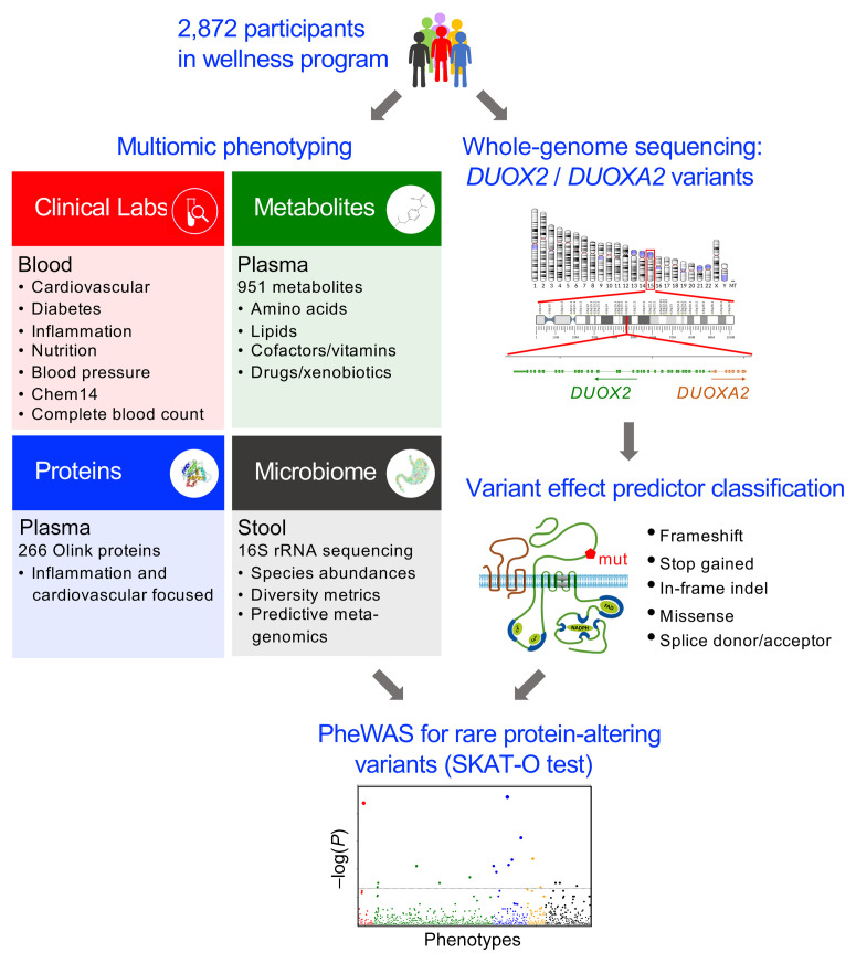
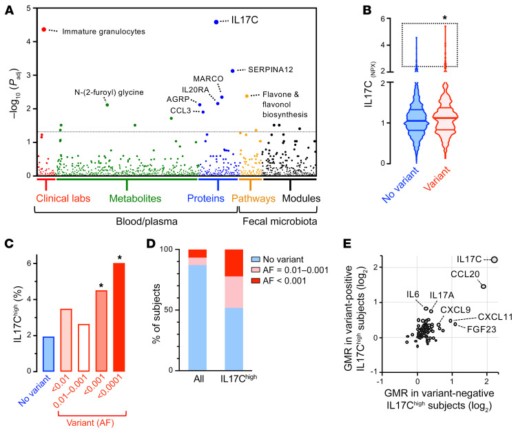
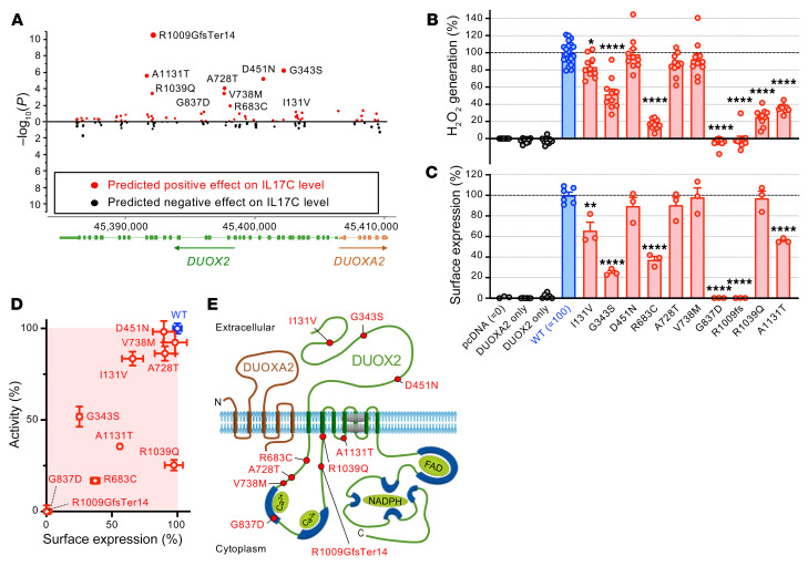
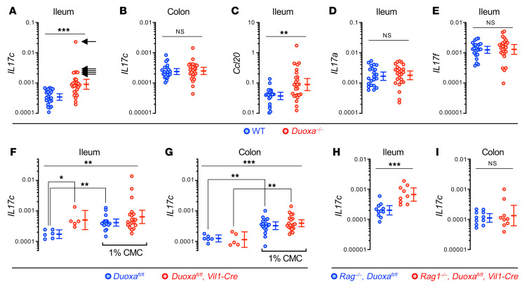
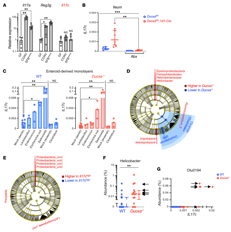
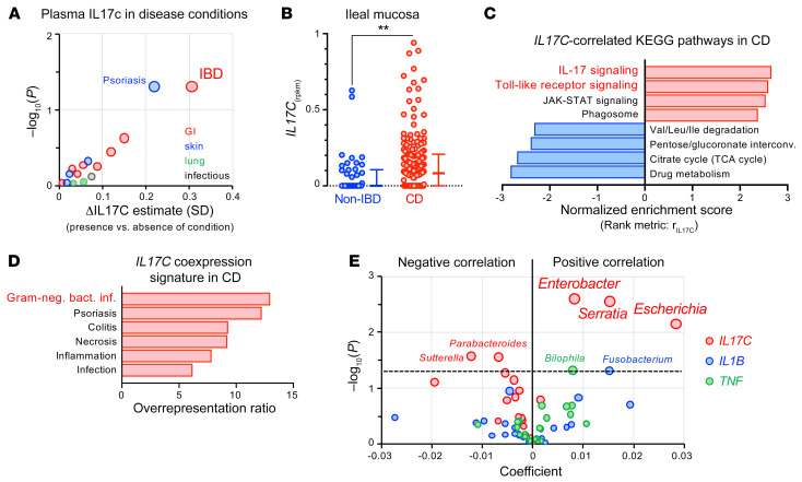
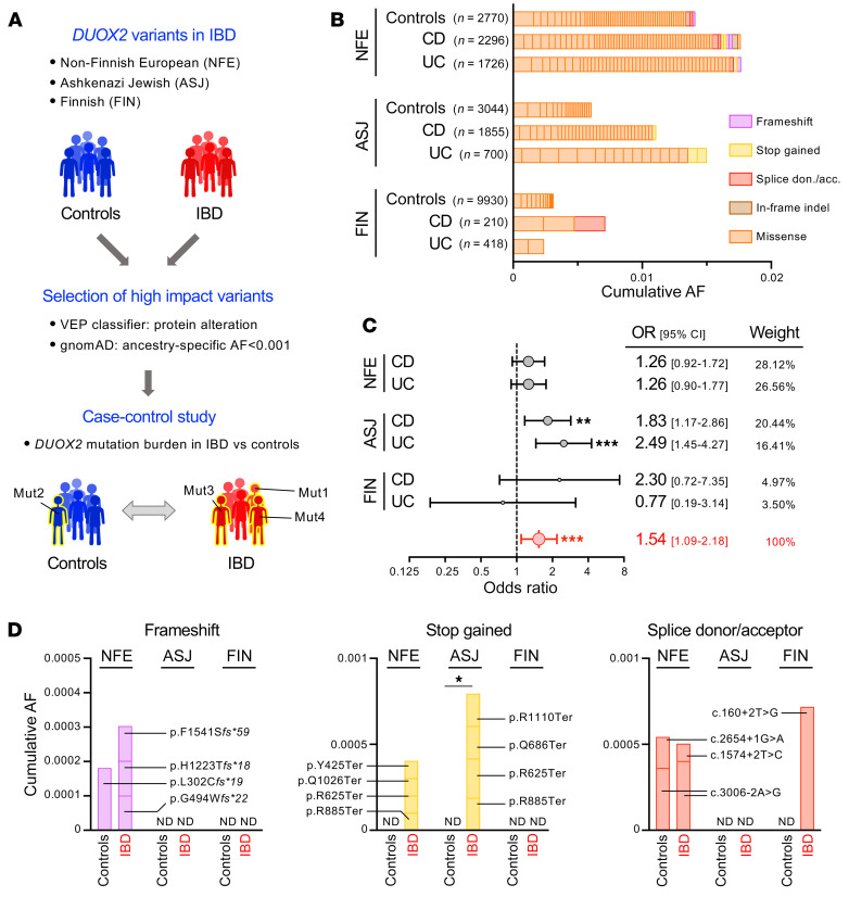
A primordial gut-epithelial innate defense response is the release of hydrogen peroxide by dual NADPH oxidase (DUOX). In inflammatory bowel disease (IBD), a condition characterized by an imbalanced gut microbiota-immune homeostasis, DUOX2 isoenzyme is the highest induced gene. Performing multiomic analyses using 2872 human participants of a wellness program, we detected a substantial burden of rare protein-altering DUOX2 gene variants of unknown physiologic significance. We identified a significant association between these rare loss-of-function variants and increased plasma levels of interleukin-17C, which is induced also in mucosal biopsies of patients with IBD. DUOX2-deficient mice replicated increased IL-17C induction in the intestine, with outlier high Il17c expression linked to the mucosal expansion of specific Proteobacteria pathobionts. Integrated microbiota/host gene expression analyses in patients with IBD corroborated IL-17C as a marker for epithelial activation by gram-negative bacteria. Finally, the impact of DUOX2 variants on IL-17C induction provided a rationale for variant stratification in case control studies that substantiated DUOX2 as an IBD risk gene. Thus, our study identifies an association of deleterious DUOX2 variants with a preclinical hallmark of disturbed microbiota-immune homeostasis that appears to precede the manifestation of IBD.

Keywords: Gastroenterology; Genetic variation; Inflammatory bowel disease; Innate immunity.


Conflict of interest statement

Conflict of interest: LAD received research funding from FrieslandCampina, Glycosyn, and Janssen.

Figures

Figure 1
Figure 1. Outline of DUOX2/DUOXA2-specific multiomic PheWAS.
We identified all rare (AF < 0.01) protein-altering DUOX2/DUOXA2 variants found in whole-genome sequencing data of 2872 participants of a lifestyle coaching program. Baseline phenotyping obtained for all participants comprised 124 clinical laboratory tests, 951 plasma metabolites, 266 plasma proteins related to inflammation and cardiovascular health, and 16S rRNA-based profiling data of the fecal microbiome. We used rare-variant test statistics (SKAT-O) to find statistical associations between the identified variants and the quantitative phenotypes.
Figure 2
Figure 2. Rare DUOX2 protein variants are associated with outlier high plasma IL-17C concentration in the general population.
(A) Manhattan plot of the PheWAS results. We used the FDR to correct for multiple testing across all combined phenotypes, with the dashed line indicating the FDR less than 0.05 significance level. (B) Plasma IL-17C baseline levels in study participants with or without DUOX2/DUOXA2 protein variants. Violin plot with quartiles indicated by the horizontal lines. Data are log2-scaled normalized protein expression units (NPX). 2-tailed Kolmogorov-Smirnov test. (C) Prevalence of high IL-17C level in subjects with or without DUOX2/DUOXA2 protein variants. We set the cut-off for outlier high IL-17C level (IL-17Chi) to Q3+2*IQR of the no-variant group and stratified variants by rarity according to ancestry-specific allele frequency (AF) data from gnomAD. Two-tailed Fisher’s exact test. (D) Enrichment of rare DUOX2 protein variants in IL-17Chi (99th percentile; n = 27) subjects. The plot depicts the proportion of individuals who carry rare DUOX2 protein variants of the indicated minor allele frequency. (E) IL-17Chi status is associated with specific alterations of the plasma protein profile that are not unique to carriers of DUOX2 protein variants. Plotted are the relative protein levels of 91 inflammation-related proteins in IL-17Chi subjects with (y axis; n = 13) or without (x axis; n = 14) rare DUOX2 protein variant(s). Protein levels are expressed as a geometric mean ratio (GMR) relative to the total study cohort. *P < 0.05.
Figure 3
Figure 3. Rare DUOX2 protein variants linked to excessive plasma IL-17C levels impair the expression of a functional DUOX2/DUOXA2 enzyme complex.
(A) Identification of variants significantly contributing to the association with plasma concentration of IL-17C in the study cohort (Wald χ2 test). (B) Extracellular H2O2 production of DUOX2 protein variants expressed in a heterologous system (9). pcDNA: transfections with empty vector; DUOXA2 only: transfections with DUOXA2 only; DUOX2 only: transfections with DUOX2 only; all other transfections are cotransfections of the indicated DUOX2 plasmids (WT or variant) with DUOXA2. Data were obtained from 3 independent transfection experiments each with 3–4 (WT: 6–8) replicates and are mean ± SEM. One-way ANOVA with Dunnett’s multiple comparisons test. (C) Quantitation of DUOX2 cell-surface expression by flow cytometry (see Supplemental Figure 2 for details). Data represent means ± SEM from 3 independent transfection experiments, each including all variants and duplicate transfections of the reference DUOX2 plasmid. One-way ANOVA with Dunnett’s multiple comparisons test. (D) Summary of the functional assessment of rare DUOX2 protein variants. Data are mean ± SEM. (E) DUOX2 topology model depicting the location of tested variants. *P < 0.05; **P < 0.01; ****P < 0.0001.
Figure 4
Figure 4. Il17c induction in the gut epithelium of DUOX2 deficient mice is T cell–independent and mimicked by impairment of the supraepithelial mucus layer.
(A and B) Il17c mRNA expression in the terminal ileum and colon of Duoxa–/– (n = 26) and WT (n = 22) littermates. Arrows indicate samples with outlier high Il17c expression (Il17chi). Ccl20 (C), Il17a (D), and Il17f (E) expression in the terminal ileum. Two-tailed Mann-Whitney. (F and G) Expression of Il17c in the ileum and colon of intestinal epithelial-specific Duoxa–/– and floxed littermate control mice. We challenged the normal bacterial compartmentalization by chronically feeding the emulsifier CMC (1% wt/vol in drinking water for 8 weeks) that thins the mucus layer (12). n = 6 and n = 17 for floxed control mice without or with CMC treatment, respectively, and n = 5 and n = 20 for intestinal epithelial-specific Duoxa–/– mice without or with CMC treatment, respectively. Kruskal-Wallis and Dunn’s post hoc test. (H and I) Il17c expression is preserved in Rag1–/– mice lacking T cells as a major source of IL-17 family cytokines. n = 11 for Rag1–/–, Duoxafl/fl mice; n = 9 for Rag1–/–, Duoxafl/fl, Vil1-Cre mice. Two-tailed Mann-Whitney. *P < 0.05; **P < 0.01; ***P < 0.001. Error bars in A–I indicate 95% CI of geometric means.
Figure 5
Figure 5. High Il17c expression in the intestinal epithelium of DUOX2-deficient mice is linked to the expansion of gram-negative pathobionts.
(A) Differential microbiota-dependent regulation of Il17c, Il17a, and Reg3g (IL-22 target gene) in the mouse intestine. GF, germ-free; CONV, conventionalized (SPF); SFBmono, monocolonized with segmented filamentous bacteria. n = 5 animals per condition. Data represent median expression values with IQR. Kruskal-Wallis with Dunn’s post hoc test. (B) Mice were treated for 3 days with an antibiotics (Abx) regimen comprising ciprofloxacin and metronidazole that suppresses the gram-negative gut microbiota (see Supplemental Figure 4). n = 6 and n = 5 for control mice without or with Abx treatment, respectively, and n = 8 and n = 4 for intestinal epithelial-specific Duoxa–/– mice without or with Abx treatment, respectively. Data represent geometric means with 95% CI. Kruskal-Wallis test with Dunn’s post hoc test. (C) Acute cell-autonomous induction of Il17c expression in enteroid-derived epithelial monolayers directly exposed to bacteria. Each treatment was performed on 6 independent enteroid cultures derived from 3 Duoxa–/–/WT littermate pairs. Bars indicate median expression values. Kruskal-Wallis with Dunn’s post hoc test. (D) Cladogram (phylum to genus level) depicting results of LEfSe (54) analysis identifying taxa with distinct relative abundance (P < 0.01; LDA > 2) in ileal mucosa of Duoxa–/– (n = 26) compared with WT (n = 22) littermates. (E) Discriminative taxa in the ileal mucosal microbiota of Il17chi animals (marked with arrows in Figure 4A). (F) The relative mucosal abundance of genus Helicobacter. Data represent median values with IQR. Two-tailed Mann-Whitney. (G) The relative abundance of Proteobacterium otu0194 vs mucosal Il17c expression. Arrows in F and G indicate animals classified as Il17chi in Figure 4A. *P < 0.05; **P < 0.01; ***P < 0.001.
Figure 6
Figure 6. IL17C induction observed in a subset of patients with IBD is a marker for abnormal epithelial stimulation by gram-negative bacteria.
(A) Positive associations of plasma IL-17C concentration with self-reported health history of study participants considering GI, skin, lung, and chronic infectious disease categories. Shown is the average difference in standardized plasma IL-17C for presence versus absence of a condition. We evaluated the nominal significance of effects using the Welch 2-sample test adjusted for age, sex, body mass index, season, and ancestry. See Supplemental Table 11 for detailed results. (B) Expression of IL17C in ileal mucosal biopsies from patients with CD (n = 174) and non-IBD controls (n = 42) from the RISK cohort. Error bars represent medians with IQR. Two-tailed Mann-Whitney. **P = 0.0027. (C) Gene set enrichment analysis using correlation with IL17C expression (rIL17C) as the rank metric to identify IL17C-correlated KEGG pathways in the mucosal biopsies of CD patients (FDR < 0.05). See Supplemental Tables 13 and 14 for additional information. (D) Overrepresentation of IL17C-coexpression signature (rIL17C > 0.5) in disease-associated gene sets from the GLAD4U database (59) (FDR < 0.05; Supplemental Table 15). (E) Multivariate association analysis using the expression of IL17C and proinflammatory cytokines (TNF, IL1B) in ileal CD biopsies (n = 135) as predictors and genus-level microbial abundance data of the mucosal microbiome as a response. Positive coefficients indicate a positive correlation between gene expression and compositional abundance of a bacterial genus (see Supplemental Tables 16 and 17 for input data and detailed results).
Figure 7
Figure 7. High impact DUOX2 variants confer increased risk for IBD.
(A) Outline of the case-control study comparing the burden of high-impact DUOX2 protein variants in patients with IBD and ancestry-matched non-IBD control cohorts. We stratified variants using population-specific allele frequencies from the gnomAD database. (B) Contribution of individual high impact DUOX2 protein variants to the cumulative allele frequencies. NFE, non–Finnish European; ASJ, Ashkenazi Jewish; FIN, Finnish. Note that the low prevalence of very rare variant carriers in Finnish participants is due to multiple genetic bottlenecks in that isolated population (23). See Supplemental Tables 18–20 for detailed data and Supplemental Figure 6 for the distribution of variants with higher allele frequencies. (C) Carriers of high-impact DUOX2 protein variants are at increased risk for developing IBD. The Forest plot depicts estimated ORs with 95% CI for patients with UC and CD from the 3 ancestry cohorts. The combined OR was calculated using a random-effects model with the Mantel-Haenszel weighting method (Supplemental Table 21). Test of the null hypothesis that OR is equal to 1 (60). (D) Detailed view of DUOX2 variants with predicted complete loss-of-function (i.e., frameshift, stop gained, and splice donor or acceptor site variants) in IBD and control cohorts. Two-tailed Fisher’s exact test. ND, not detected. *P < 0.05; **P < 0.01; ***P < 0.001.

Comment in

References

    1. Ha EM, et al. A direct role for dual oxidase in Drosophila gut immunity. Science. 2005;310(5749):847–850. doi: 10.1126/science.1117311. - DOI - PubMed
    1. Chavez V, et al. Ce-Duox1/BLI-3 generates reactive oxygen species as a protective innate immune mechanism in Caenorhabditis elegans. Infect Immun. 2009;77(11):4983–4989. doi: 10.1128/IAI.00627-09. - DOI - PMC - PubMed
    1. Grasberger H, et al. Dual oxidases control release of hydrogen peroxide by the gastric epithelium to prevent Helicobacter felis infection and inflammation in mice. Gastroenterology. 2013;145(5):1045–1054. doi: 10.1053/j.gastro.2013.07.011. - DOI - PMC - PubMed
    1. Collins KD, et al. Chemotaxis allows bacteria to overcome host-generated reactive oxygen species that constrain gland colonization. Infect Immun. 2018;86(5):e00878-17. - PMC - PubMed
    1. Grasberger H, et al. Increased expression of DUOX2 Is an epithelial response to mucosal dysbiosis required for immune homeostasis in mouse intestine. Gastroenterology. 2015;149(7):1849–1859. doi: 10.1053/j.gastro.2015.07.062. - DOI - PMC - PubMed

Publication types