. 2021 Mar 2;15(3):e0009230.
doi: 10.1371/journal.pntd.0009230. eCollection 2021 Mar.

Protein methyltransferase 7 deficiency in Leishmania major increases neutrophil associated pathology in murine model

Affiliations

Protein methyltransferase 7 deficiency in Leishmania major increases neutrophil associated pathology in murine model

Juliana Alcoforado Diniz et al. PLoS Negl Trop Dis. .

Abstract

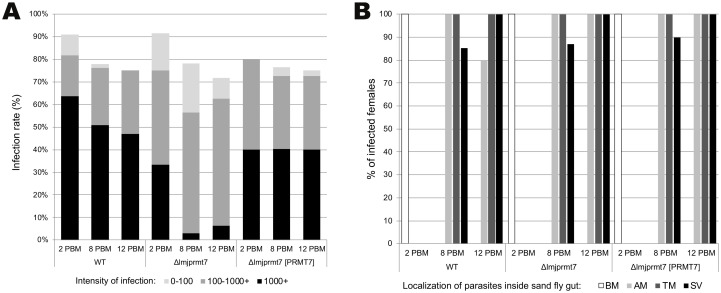
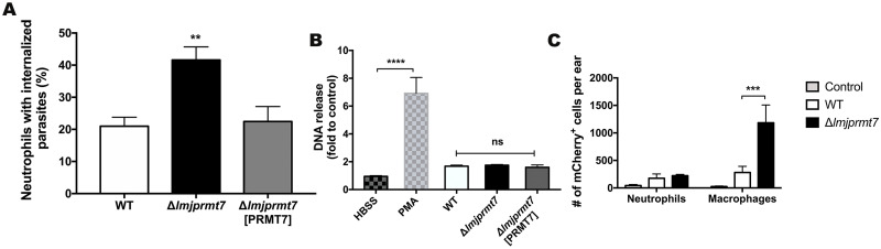
Leishmania major is the main causative agent of cutaneous leishmaniasis in the Old World. In Leishmania parasites, the lack of transcriptional control is mostly compensated by post-transcriptional mechanisms. Methylation of arginine is a conserved post-translational modification executed by Protein Arginine Methyltransferase (PRMTs). The genome from L. major encodes five PRMT homologs, including the cytosolic protein associated with several RNA-binding proteins, LmjPRMT7. It has been previously reported that LmjPRMT7 could impact parasite infectivity. In addition, a more recent work has clearly shown the importance of LmjPRMT7 in RNA-binding capacity and protein stability of methylation targets, demonstrating the role of this enzyme as an important epigenetic regulator of mRNA metabolism. In this study, we unveil the impact of PRMT7-mediated methylation on parasite development and virulence. Our data reveals that higher levels of LmjPRMT7 can impair parasite pathogenicity, and that deletion of this enzyme rescues the pathogenic phenotype of an attenuated strain of L. major. Interestingly, lesion formation caused by LmjPRMT7 knockout parasites is associated with an exacerbated inflammatory reaction in the tissue correlated with an excessive neutrophil recruitment. Moreover, the absence of LmjPRMT7 also impairs parasite development within the sand fly vector Phlebotomus duboscqi. Finally, a transcriptome analysis shed light onto possible genes affected by depletion of this enzyme. Taken together, this study highlights how post-transcriptional regulation can affect different aspects of the parasite biology.


Conflict of interest statement

The authors have declared that no competing interests exist.

Figures

Fig 1
Fig 1. Deletion of LmjPRMT7 results in a decrease in the number of parasites inside the P. duboscqi vector.
(A) Infection rates and parasite load in P. duboscqi on day 2, 8 and 12 post blood meal. Abbreviations: 0 no infection; 0–100 low infection; 100–1000 medium infection; 1000+ heavy infection. (B) Localization of parasites inside the P. duboscqi gut on day 2, 8 and 12 post blood meal. Abbreviations: SV—stomodeal valve, TM—thoracic midgut, AM—abdominal midgut, BM—within the bloodmeal. Data originate from 4 independent experiments; sand flies were infected by 106 parasites per ml of bloodmeal.
Fig 2
Fig 2. Higher levels of PRMT7 enzyme are found in a nonpathogenic strain of L. major.
(A) Quantitative RT-PCR analysis of RNA extracted from promastigotes during the early-log (procyclic) culture phase and purified metacyclic. Levels of PRMT7 were comparable between the pathogenic Leishmania major strains: Ryan, LV39, Sd and Friedlin (Fn) in comparison to the nonpathogenic CC1 strain. The expression is relative to the expression of the G6PD and RNA45 genes. The results are shown as mean ± SEM. p < 0.05 (*) and p < 0.01 (**) were considered statistically significant. Statistical analysis was performed by Two-way ANOVA followed by Sidak post-test. (B) BALB/c mice (n = 5 per group) were injected with 105 metacyclic promastigotes of the different strains of L. major and ear thicknesses were followed weekly. (C) Image of infected ears 5 weeks post infection and photos were taken by the author. Five weeks after infection, parasite titers were determined in the ear (D) and draining lymph node (E). The results are shown as mean ± SEM. For the parasite burden, statistical analysis was conducted comparing virulent strains with the LmjCC1 attenuated line. Statistical analysis was performed by unpaired, two-tailed student’s t-test. *p<0.05.
Fig 3
Fig 3. Deletion of LmjPRMT7 in a nonpathogenic strain leads to lesion formation not affecting parasite load.
(A) BALB/c female mice (n = 5 per group) were injected intradermally in both ears with 100,000 metacyclic promastigotes and ear thicknesses was followed for 7 weeks. (B) Photographs of infected ears on the 7th week post infection, taken by the author. Results shown are the mean ± SEM of twenty ears, five mice/group, from two independent experiments, **p < 0.01, ****p<0.0001, comparing infection with wild type and Δlmjprmt7, using 2way ANOVA, with Tukey’s multiple comparison test. (C-D) Parasite burden determined by limiting dilution analysis (LDA) in the site of infection (ear, left) and draining lymph node (LN, right) after 7 weeks post-infection. Results shown are the mean ± SEM of fourteen ears from two independent experiments. Unpaired, two-tailed student’s t-test.
Fig 4
Fig 4. Subset of myeloid cells recruited to the site of infection after 7 weeks post-infection.
BALB/c mice were infected in the ear dermis with 100,000 Lmj CC1 metacyclic promastigotes from the wild type, Δlmjprmt7 and Δlmjprmt7 [PRMT7] strains. Ear tissues were processed at 7 weeks post-infection to study the cell recruitment by flow cytometry. (A) Representative dot plots of ear-derived dermal cells. Subpopulations of myeloid (CD11b+) cells are defined as 1. Neutrophils, 2. Eosinophils, 3. Conventional dendritic cells, 4. Dermal macrophages, 5. Monocyte derived dendritic cells, 6. Inflammatory monocytes. (B) The number of cells in each myeloid cell subset after 7 weeks of infection (NΦ: neutrophils; Eos: eosinophils; DCs: dendritic cells; Dermal MΦ: dermal macrophages; Infl. MNC: inflammatory monocytes; MNC DCs: Monocyte derived dendritic cells). All data are shown as the mean ± SEM of twenty samples (five mice, per group) from two independent experiments ***p<0.001, using ordinary one-way ANOVA, with Dunn’s multiple comparison test. (C) The representative contour plot of another independent experiment and (D) the percentages of neutrophils (CD11b+Ly6G+ cells) after 7 weeks of infection.
Fig 5
Fig 5. Infection of resistant and susceptible mice strains display similar lesion outcome.
(A) C57BL/6 and (B) BALB/c mice were injected in the ear with 105 metacyclic promastigotes of Lmj CC1 WT and transfectants (n = 6 mice per group), and the ear thicknesses were followed for 7 weeks. Images of infected ears are shown next to each graph as follow: 1 WT, 2 ΔlmjPRMT7 and 3 ΔlmjPRMT7 [PRMT7] and pictures were taken by the author. (C-D) Parasite quantification at 7 weeks post-infection. The results are shown as mean ± SEM. Statistical analysis was performed by unpaired, two-tailed student’s t-test.
Fig 6
Fig 6. Neutrophils display increased uptake of parasites lacking PRMT7 in in vitro assays.
(A) To evaluate the uptake of parasites by neutrophils, 10,000 BMDN were incubated for 3 hours with metacyclic promastigotes (MOI 1). The cells were centrifuged on slides and stained for evaluation by light microscopy. Data are expressed as the mean of neutrophils with intracellular parasites ± SEM. ** p<0.01. Statistical analysis was performed by ordinary one-way ANOVA followed by Tukey’s multiple comparison test. These results are a pool of 3 independent experiments. (B) NET formation was assessed after incubating 10,000 BMDNs with modified strains of L. major CC1, PMA (50nM), or medium (untreated) at MOI of 1 for 3 hours. DNA release after the treatment/infection was quantified in the cell supernatants by fluorescence detection (Ex./Em. 480/520 nm) after reaction with SYTOX green. Data are expressed as a mean of fluorescence intensity ± SEM; **** p<0.0001. Statistical analysis was performed by two-way ANOVA followed by Tukey’s multiple comparison test. DNA quantitation assays were performed in duplicate and, the data shown are the pool of two independent experiments. (C) To evaluate the uptake in vivo by flow cytometry, 4x10⁵ Lmj CC1 metacyclic promastigotes from the wild type and Δlmjprmt7 fluorescently tagged with mCherry were used to infect BALB/c mice. Ear tissues were processed at 4-hours post-infection to study the number of macrophages and neutrophils infected with the different fluorescent strains. Data are shown as the mean ± SEM of four samples per group. Representative data from two independent experiments. *** p<0.001, using two-way ANOVA followed by Tukey’s multiple comparison test.
Fig 7
Fig 7. Analysis of transcripts differentially expressed in the LmjPRMT7 compared transfectants.
(A) Venn diagram of the three contrasts obtained after differentially expressed analysis of transcriptomes from L. major parasite samples (WT, ΔlmjPRMT7 and ΔlmjPRMT7 [PRMT7]). In bold the DEG only differentially expressed in Δ7 compared to WT and AB but not between the last two groups (FDR 5%, FC > 1.5). On the right bottom corner, the number of genes with no expression difference found between the groups. (B) Description of genes showed in bold in the Venn Diagram. (C) The convergent switch strand region on chromosome 28 depicting the location of three of the DEG identified, LmjF.28.1580, LmjF.28.1590 and LmjF.28.1570.

Similar articles

Cited by

References

    1. Georgiadou SP, Makaritsis KP, Dalekos GN. Leishmaniasis revisited: Current aspects on epidemiology, diagnosis and treatment. J Transl Intern Med. 2016. 10.1515/jtim-2015-0002 - DOI - PMC - PubMed
    1. Akhoundi M, Kuhls K, Cannet A, Votýpka J, Marty P, Delaunay P, et al.. A Historical Overview of the Classification, Evolution, and Dispersion of Leishmania Parasites and Sandflies. PLoS Neglected Tropical Diseases. 2016. 10.1371/journal.pntd.0004349 - DOI - PMC - PubMed
    1. Sacks D, Noben-Trauth N. The immunology of susceptibility and resistance to Leishmania major in mice. Nat Rev Immunol. 2002. 10.1038/nri933 - DOI - PubMed
    1. Ready PD. Biology of Phlebotomine Sand Flies as Vectors of Disease Agents. Annu Rev Entomol. 2013. 10.1146/annurev-ento-120811-153557 - DOI - PubMed
    1. Dostálová A, Volf P. Leishmania development in sand flies: Parasite-vector interactions overview. Parasites and Vectors. 2012. 10.1186/1756-3305-5-276 - DOI - PMC - PubMed

Publication types

MeSH terms

Substances