Review
. 2021 May 6:50:447-468.
doi: 10.1146/annurev-biophys-062920-063555. Epub 2021 Mar 2.

Measuring Absolute Membrane Potential Across Space and Time

Affiliations
Review

Measuring Absolute Membrane Potential Across Space and Time

Julia R Lazzari-Dean et al. Annu Rev Biophys. .

Abstract

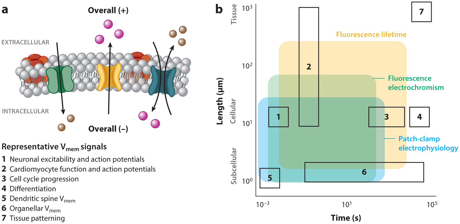
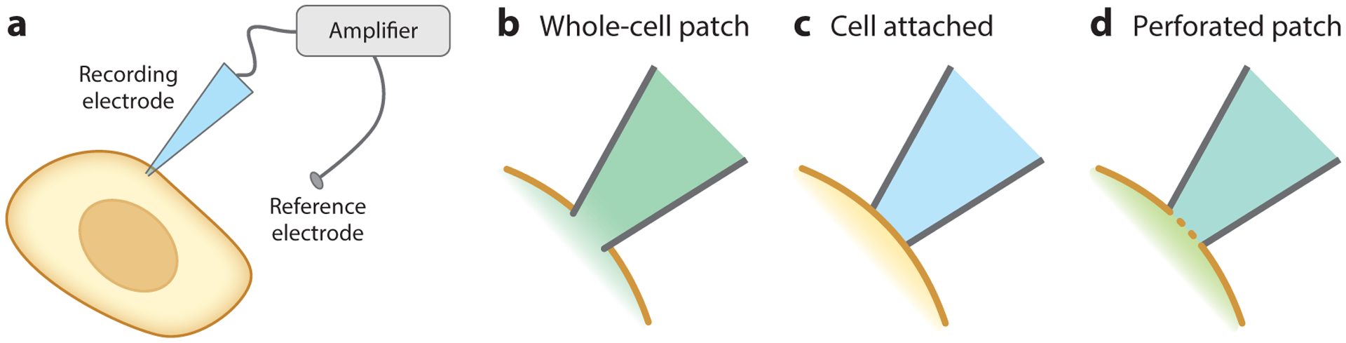
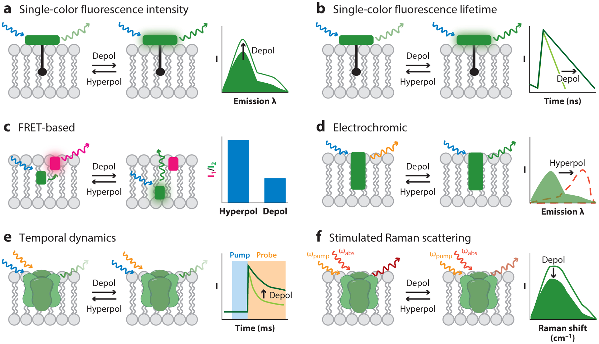
Membrane potential (Vmem) is a fundamental biophysical signal present in all cells. Vmem signals range in time from milliseconds to days, and they span lengths from microns to centimeters. Vmem affects many cellular processes, ranging from neurotransmitter release to cell cycle control to tissue patterning. However, existing tools are not suitable for Vmem quantification in many of these areas. In this review, we outline the diverse biology of Vmem, drafting a wish list of features for a Vmem sensing platform. We then use these guidelines to discuss electrode-based and optical platforms for interrogating Vmem. On the one hand, electrode-based strategies exhibit excellent quantification but are most effective in short-term, cellular recordings. On the other hand, optical strategies provide easier access to diverse samples but generally only detect relative changes in Vmem. By combining the respective strengths of these technologies, recent advances in optical quantification of absolute Vmem enable new inquiries into Vmem biology.

Keywords: electrophysiology; fluorescence; fluorescence lifetime; membrane potential; microscopy; quantitative imaging.


Figures

Figure 1
Figure 1
Membrane potential (Vmem) signals and measurement techniques across time and space. (a) A schematic of cellular Vmem, with examples of seven biological processes where Vmem signaling plays a role. The resistance to ionic flow between compartments sets the length scale of Vmem signals; Vmem can scale across tissues or be compartmentalized within organelles or dendritic spines. The properties of ion channels and pumps that determine Vmem, as well as ion diffusion between compartments, dictate the time duration of a Vmem response. (b) Biological space accessible to the three most common strategies for absolute Vmem measurement (shaded regions), overlaid with example biological process of interest (outlined boxes, numbered as in panel a). While many techniques can report cellular Vmem on the scale of milliseconds or seconds, longer recordings or recordings across large areas are difficult to access. Shaded areas indicate regions where single trial recordings retain voltage resolution on the scale of biological Vmem changes (≤20 mV) and minimally alter the biological sample. Recordings with fluorescence electrochromic dyes require recalibration with an electrode on each cell, whereas fluorescence lifetime and patch-clamp electrophysiology can be applied as stand-alone measurements that are comparable between cells. Shaded areas are restricted to demonstrated application space for each technique and do not indicate the full extent of its possible use.
Figure 2
Figure 2
Electrophysiological configurations for Vmem recording. (a) General schematic for electrode-based Vmem recordings, in which a cell comes into direct contact with an electrode. Voltage is measured as the difference between the recording electrode and a reference electrode in the bath solution. (bd) Close-up of the interface between the membrane and the electrode in different electrophysiology configurations. (b) In whole-cell patch-clamp electrophysiology, the plasma membrane is ruptured, and the cytosol mixes with the recording electrode solution. (c) In the cell-attached configuration, the plasma membrane is left intact. (d) In perforated patch, the membrane is not ruptured, but ionophores introduced into the recording solution allow ionic exchange across the membrane.
Figure 3
Figure 3
Optical strategies for reporting absolute Vmem. (a) Single-color fluorescence intensity recordings detect changes in Vmem by changes in a sensor’s fluorescence quantum yield (shown), extinction coefficient, or cellular concentration. This technique generally cannot report absolute Vmem optically, although estimates can be made if calibrations are performed for every cell of interest. (b) Single-color fluorescence lifetime can report absolute Vmem, particularly if the Vmem sensor operates via photoinduced electron transfer.(c) FRET-based sensors show differences in the FRET ratios of two fluorophores, often resulting from Vmem-dependent changes in the distance between the two. The diagram shows a FRET-oxonol system. (d) The excitation and/or emission spectra of electrochromic dyes depend on the electric field in the plasma membrane. (e) Certain GEVIs show Vmem-related differences in the temporal dynamics of the absorption of their excited state. (f) SRS imaging reports changes in Vmem-dependent vibrational frequencies. Abbreviations:λ, wavelength; depol, depolarization; FRET, Förster resonance energy transfer; GEVI, genetically encoded voltage indicator; hyperpol, hyperpolarization; SRS, stimulated Raman scattering; Vmem, membrane potential.

References

    1. Abdelfattah AS, Kawashima T, Singh A, Novak O, Liu H, et al. 2019. Bright and photostable chemigenetic indicators for extended in vivo voltage imaging. Science 365(6454):699–704 - PubMed
    1. Abdul Kadir L, Stacey M, Barrett-Jolley R. 2018. Emerging roles of the membrane potential: action beyond the action potential. Front. Physiol 9:1661. - PMC - PubMed
    1. Adam Y, Kim JJ, Lou S, Zhao Y, Xie ME, et al. 2019. Voltage imaging and optogenetics reveal behaviour-dependent changes in hippocampal dynamics. Nature 569:413–17 - PMC - PubMed
    1. Akemann W, Mutoh H, Perron A, Park YK, Iwamoto Y, Knopfel T. 2012. Imaging neural circuit dynamics with a voltage-sensitive fluorescent protein. J. Neurophysiol 108(8):2323–37 - PubMed
    1. Alcamí P, Pereda AE. 2019. Beyond plasticity: the dynamic impact of electrical synapses on neural circuits. Nat. Rev. Neurosci 20(5):253–71 - PubMed

Publication types