. 2021 Mar 2;135(4):1026-1035.
doi: 10.3171/2020.8.JNS201810. Print 2021 Oct 1.

Second window ICG predicts gross-total resection and progression-free survival during brain metastasis surgery

Affiliations

Second window ICG predicts gross-total resection and progression-free survival during brain metastasis surgery

Clare W Teng et al. J Neurosurg. .

Abstract

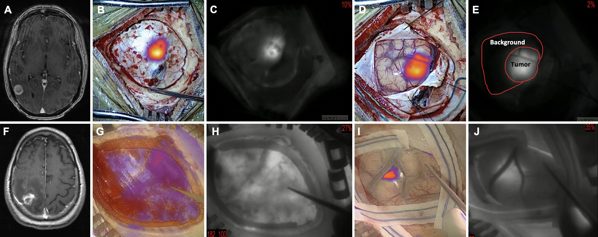
Objective: Metastases are the most common intracranial malignancies and complete resection can provide relief of neurological symptoms and reduce recurrence. The authors' prospective pilot study in 2017 demonstrated promising results for the application of high-dose, delayed imaging of indocyanine green (ICG), known as second window ICG (SWIG), in patients undergoing surgery for brain metastases. In this prospective cohort study, the authors evaluated intraoperative imaging and clinical outcomes of treatment using SWIG.

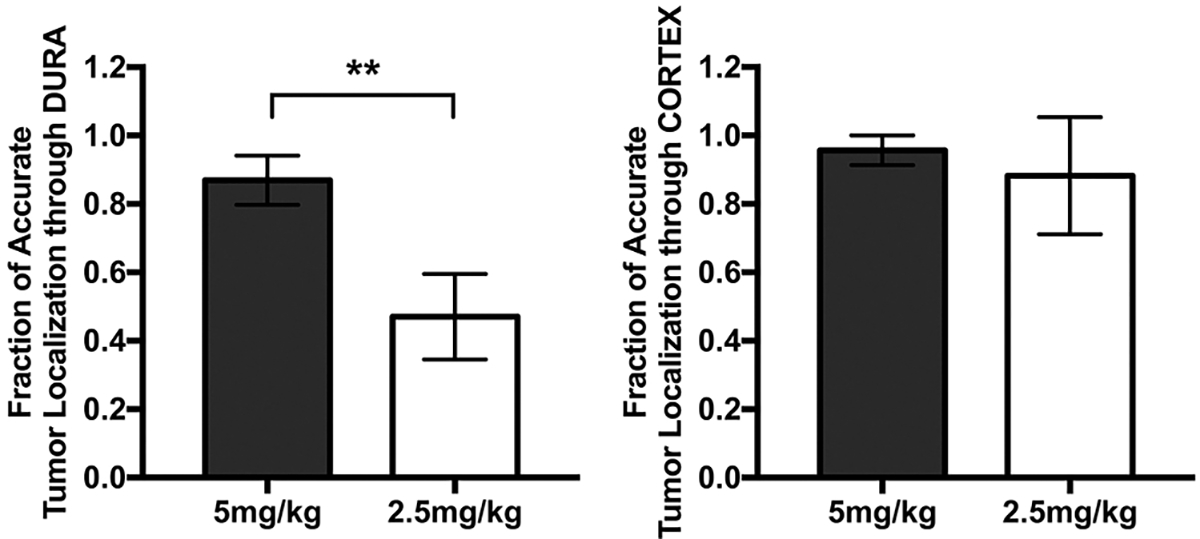
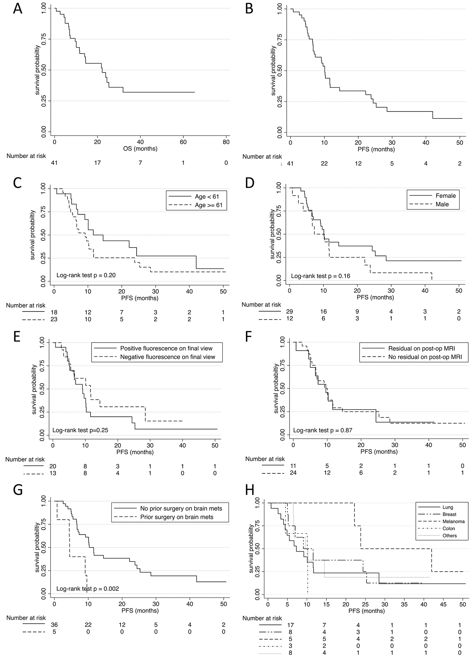
Methods: Patients were prospectively enrolled in an approved study of high-dose, delayed ICG (SWIG) and received 5 mg/kg (2014-2018) or 2.5 mg/kg (2018-2019) ICG 24 hours preoperatively. Intraoperatively, near-infrared (NIR) imaging was performed using a dedicated NIR exoscope. NIR images were analyzed and the signal-to-background ratio (SBR) was calculated to quantify fluorescence. Residual fluorescence on the postresection NIR view was compared and correlated to the residual gadolinium enhancement on postoperative MRI. Patient survival and predictive factors were analyzed.

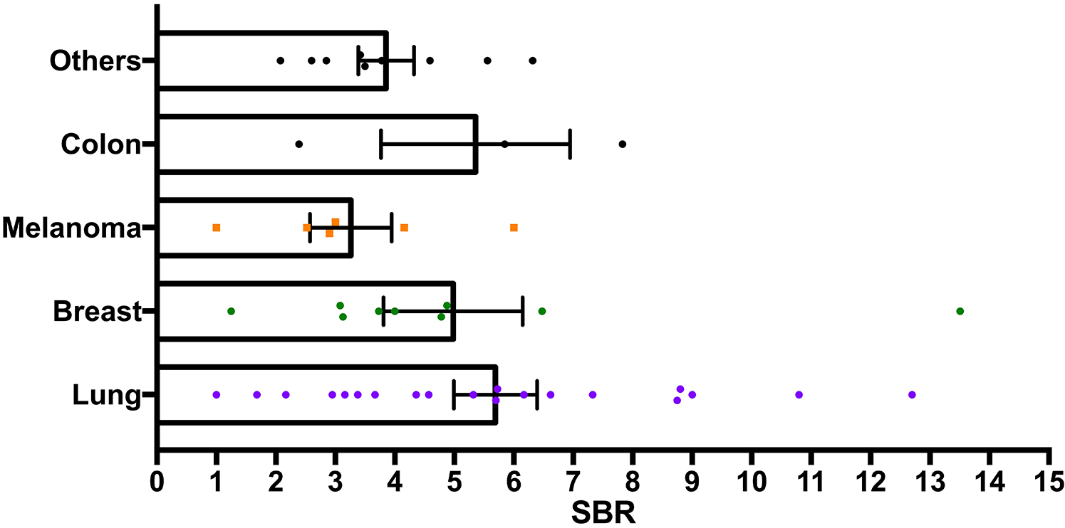
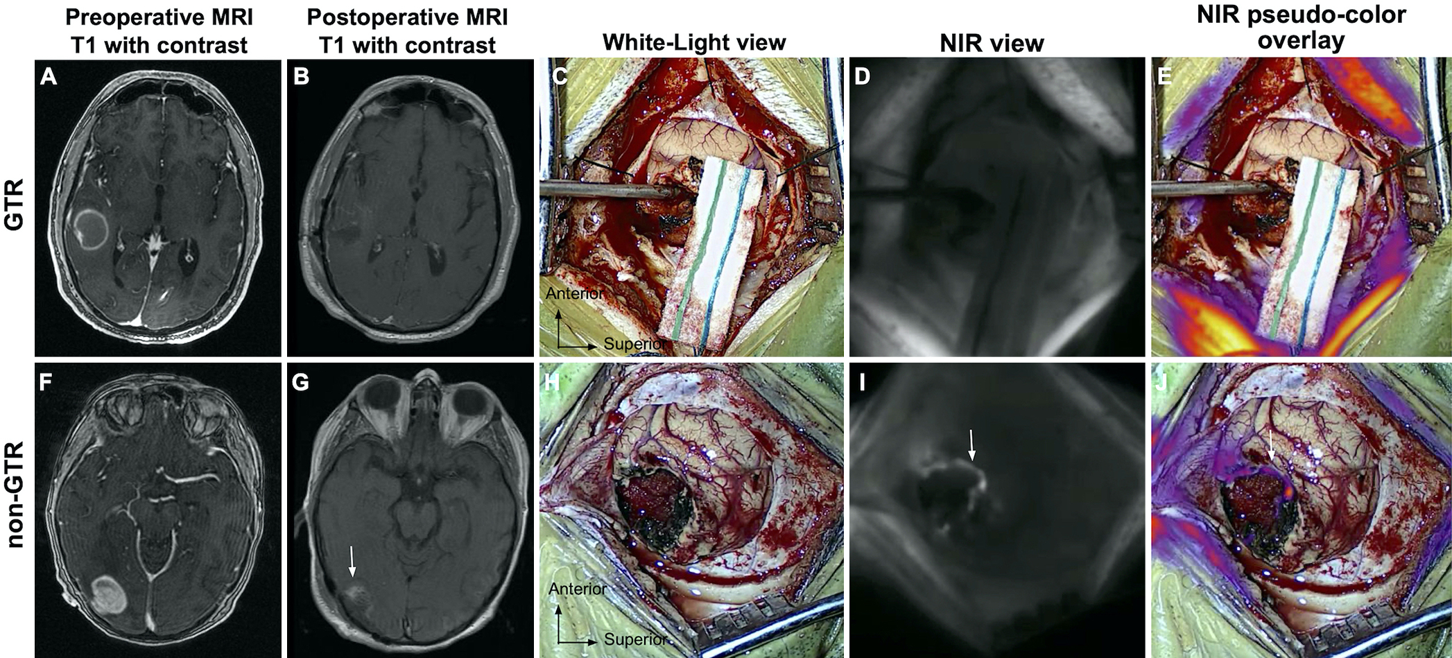
Results: In total, 51 intracranial metastases were surgically treated in 47 patients in this cohort. All 51 metastatic tumors demonstrated strong NIR fluorescence (mean SBR 4.9). In tumors ≤ 10 mm from the cortical surface, SWIG with 5 mg/kg ICG produced enhanced transdural tumor visibility (91.3%) compared to 2.5 mg/kg (52.9%; p = 0.0047). Neoplastic margin detection using NIR fluorescence compared to white light improved sensitivity, albeit lowered specificity; however, increasing the SBR cutoff for positive fluorescence significantly improved specificity without sacrificing sensitivity, increasing the overall accuracy from 57.5% to 72.5%. A lack of residual NIR fluorescence after resection was closely correlated with a lack of residual enhancement on postoperative MRI (p = 0.007). Among the 16 patients in whom tumor recurred at the site of surgery, postoperative MRI successfully predicted 8 cases, whereas the postresection NIR view predicted 12 cases. Progression-free survival rate at 12 months was greater for patients without residual NIR fluorescence (38%) than for those without residual enhancement on postoperative MRI (29%).

Conclusions: The current study demonstrates the clinical benefits of the SWIG technique in surgery for patients with brain metastases. Specifically, this technique allows for dose-dependent, transdural localization of neoplasms and improved sensitivity in neoplastic margin detection. Postresection residual fluorescence can be a powerful tool to evaluate extent of resection in conjunction with MRI, and it may guide decisions on brain metastasis management.

Keywords: SWIG; brain metastasis; near-infrared fluorescence; oncology; postoperative MRI; recurrence; second window indocyanine green.


Figures

FIG. 1.
FIG. 1.
Examples of successful (5 mg/kg) and unsuccessful (2.5 mg/kg) tumor localization through dura/cortex. A–E: The patient was infused intravenously with 5 mg/kg ICG approximately 24 hours before surgery. T1 MRI was performed with gadolinium at 1-mm resolution. The axial image (A) demonstrated a contrast-enhancing superficial mass extending to the cortex, consistent with a metastasis. Intraoperatively, after craniotomy and prior to dura opening, NIR fluorescence was visualized in both pseudocolor overlay (B) and black and white (C), demonstrating successful visualization of the tumor through the intact dura. After durotomy, the NIR cortex view was imaged (D and E) and the localization of tumor on the dura view and cortex view was consistent (E). Demonstration of the method of SBR calculation: signal = 92.7, background = 9.2, SBR = 10.1. F–J: The patient was infused intravenously with 2.5 mg/kg ICG approximately 24 hours before surgery. T1 MRI was performed with gadolinium at 1-mm resolution. The axial image (F) demonstrated a contrast-enhancing superficial mass extending to the cortex, consistent with a metastasis. Intraoperatively, after craniotomy and prior to dura opening, the NIR fluorescence image was visualized (G and H). The location of the tumor was unable to be determined solely based on the dura view. After durotomy, the NIR cortex view was visualized. In this case, the location of the tumor was successfully visualized on the cortex view. Some nonspecific signal from the skull is seen in the lower-left corner (I and J). Figure is available in color online only.
FIG. 2.
FIG. 2.
Fractions of tumor accurately localized through the dura and cortex. Left: NIR signal on dura view was evaluated for its ability to guide localization of the tumor. The localization was deemed successful by the independent reviewer if the location of the tumor determined on the dura view corresponded with the location shown on the tumor view. A statistically significant difference in the fraction of accurate tumor localization through dura between the two dose groups was demonstrated (**p = 0.0047). Bars represent the standard error. Right: Postdurotomy, a similar analysis was performed on the cortex view. The difference between the two groups was not statistically significant (p = 0.39). Bars represent the standard error.
FIG. 3.
FIG. 3.
Pathology-based SBR comparison: comparison of average SBRs of metastases of lung cancer, melanoma, breast cancer, colon cancer, and other pathologies. A significantly lower SBR of the melanoma pathologies (p = 0.047) was demonstrated; NIR fluorescence from the other tumors was not statistically different. Figure is available in color online only.
FIG. 4.
FIG. 4.
Correlation between postresection NIR view with postoperative MRI. A–E: Absence of NIR fluorescence on NIR final view predicts complete resection of the enhancing tumor on postoperative MRI. Preoperative T1 MRI with gadolinium contrast (A) demonstrating a 3.1 × 2.9–cm contrast-enhancing intracranial metastasis in the right temporal lobe. Postoperative T1 MRI with gadolinium contrast (B) demonstrated GTR with minimal peripheral enhancement consistent with postoperative changes. Both WL (C) and NIR (D and E) imaging of the surgical cavity after resection suggested the absence of residual neoplasm. F–J: Residual fluorescence on the NIR final view corresponds with residual contrast-enhancing tissue on postoperative MRI. Preoperative T1 MRI with gadolinium contrast (F) demonstrating a 3.0 × 2.5–cm contrast-enhancing intracranial metastasis in the right occipital lobe. Postoperative T1 MRI with gadolinium contrast (G) demonstrated residual nodular enhancement on the anterior surface of the resection cavity. The postresection WL view (H) did not show evidence of residual neoplasm. NIR final view imaging revealed residual fluorescence with nodular appearance along the anterior resection cavity, corresponding with the findings on MRI (I and J). Figure is available in color online only.
FIG. 5.
FIG. 5.
Kaplan-Meier survival curves. A: OR was 19 months (range 0.7–65.3 months). B: PFS was 15 months (range 0.7–50.7 months). C–H: PFS stratified by age (C), sex (D), NIR final view (E), postoperative MRI (F), prior surgery on BM (G), and primary pathology (H). mets = metastases.

References

    1. Dagogo-Jack I, Carter SL, Brastianos PK. Brain metastasis: clinical implications of branched evolution. Trends Cancer. 2016;2(7): 332–337. - PubMed
    1. Ferguson SD, Wagner KM, Prabhu SS, et al. Neurosurgical management of brain metastases. Clin Exp Metastasis. 2017; 34(6–7): 377–389. - PubMed
    1. Lowery FJ, Yu D. Brain metastasis: unique challenges and open opportunities. Biochim Biophys Acta Rev Cancer. 2017; 1867(1): 49–57. - PMC - PubMed
    1. Nayak L, Lee EQ, Wen PY. Epidemiology of brain metastases. Curr Oncol Rep. 2012; 14(1): 48–54. - PubMed
    1. Prabhu RS, Miller KR, Asher AL, et al. Preoperative stereotactic radiosurgery before planned resection of brain metastases: updated analysis of efficacy and toxicity of a novel treatment paradigm. J Neurosurg. 2019;131(5):1387–1394. - PubMed

LinkOut - more resources