. 2021 Feb 26;11(3):395.
doi: 10.3390/diagnostics11030395.

Preclinical Evaluation of Artesunate as an Antineoplastic Agent in Ovarian Cancer Treatment

Affiliations

Preclinical Evaluation of Artesunate as an Antineoplastic Agent in Ovarian Cancer Treatment

Anthony McDowell Jr et al. Diagnostics (Basel). .

Abstract

Background: Ovarian cancer is the deadliest gynecologic malignancy despite current first-line treatment with a platinum and taxane doublet. Artesunate has broad antineoplastic properties but has not been investigated in combination with carboplatin and paclitaxel for ovarian cancer treatment.

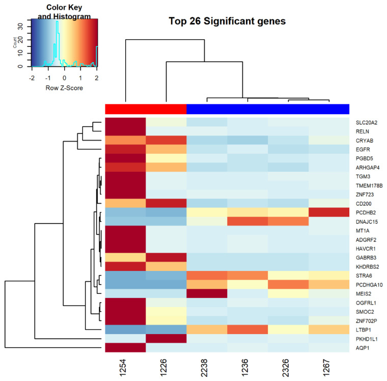
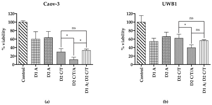
Methods: Standard cell culture technique with commercially available ovarian cancer cell lines were utilized in cell viability, DNA damage, and cell cycle progression assays to qualify and quantify artesunate treatment effects. Additionally, the sequence of administering artesunate in combination with paclitaxel and carboplatin was determined. The activity of artesunate was also assessed in 3D organoid models of primary ovarian cancer and RNAseq analysis was utilized to identify genes and the associated genetic pathways that were differentially regulated in artesunate resistant organoid models compared to organoids that were sensitive to artesunate.

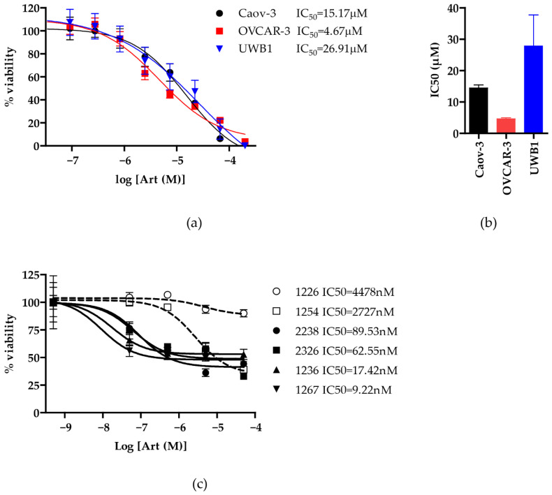
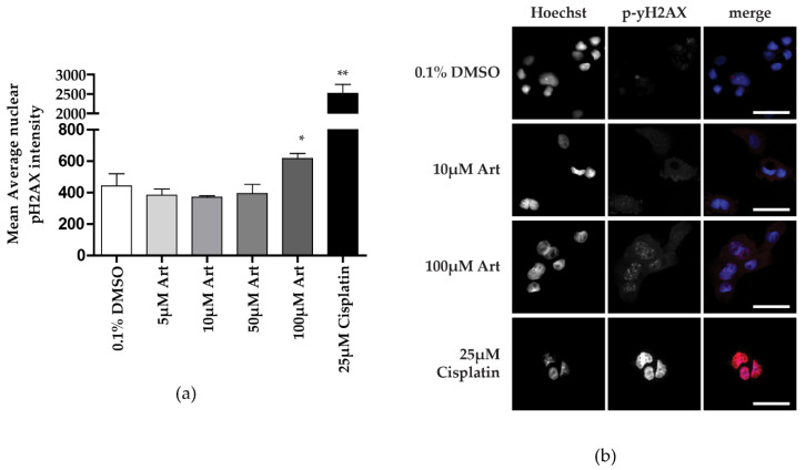
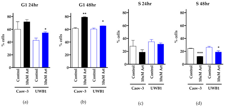
Results: Artesunate treatment reduces cell viability in 2D and 3D ovarian cancer cell models. Clinically relevant concentrations of artesunate induce G1 arrest, but do not induce DNA damage. Pathways related to cell cycle progression, specifically G1/S transition, are upregulated in ovarian organoid models that are innately more resistant to artesunate compared to more sensitive models. Depending on the sequence of administration, the addition of artesunate to carboplatin and paclitaxel improves their effectiveness.

Conclusions: Artesunate has preclinical activity in ovarian cancer that merits further investigation to treat ovarian cancer.

Keywords: Artemesia annua; artesunate; carboplatin; dihydroartemisinin; ovarian cancer; paclitaxel.


Conflict of interest statement

The authors declare no conflict of interest.

Figures

Figure 1
Figure 1
Artesunate sensitivities across 3 commercially available ovarian cancer cell lines and 6 novel 3D ovarian cancer organoids. (a) Human ovarian cancer cell lines Caov-3, OVCAR-3, and UWB1.289 cells were treated with serially diluted concentrations of artesunate for 72 h. CellTiter-Glo 2.0 viability assay (Promega) was used to calculate percent viability from the proliferation of treated versus vehicle-treated control cells. The mean +/− SD from three independent experiments is shown graphically and IC50s were calculated using a variable slope non-linear regression line. (b) The mean IC50 of artesunate treatments in each cell line is graphed with SD. Utilizing a one-way ANOVA with Bonferroni’ Multiple Comparison Test, all IC50s were not significantly different with p > 0.05. (c) 3D ovarian organoids were established from 6 patients and were treated similarly with serially diluted concentrations of artesunate for 72 h. Viability was measured with MTS assay (Promega) and the signal for each drug treatment was normalized to a matched vehicle-treated control. The mean % viability +/− SD is shown graphically and IC50s were calculated using a variable slope non-linear regression line.
Figure 2
Figure 2
Heat map showing the top 26 most significantly differentially expressed genes between ovarian organoid models that are resistant to artesunate (1254 and 1226) compared to models that are sensitive to artesunate (2238, 2326, 1267, and 1236). The red bar along the top of the heat map denotes the resistant models while the blue bar indicates the sensitive models. RNAseq analysis was performed on RNA extracted from untreated cells.
Figure 3
Figure 3
DNA damage assay as average nuclear pH2AX intensity. (a) The average nuclear intensity of pH2AX staining was quantified in Caov-3 cells treated for 48 h with artesunate concentrations ranging from 5–100 μM with 25 μM cisplatin treatment as a positive control. The mean signal +/− SD was graphed and a one-tailed t-test was performed (* p = 0.0486, ** p = 0.0034) (b) Representative images of cells treated with 0.1% DMSO (vehicle), 10 μM artesunate, 100 μM artesunate, or 25 μM cisplatin (positive control for DNA damage).
Figure 4
Figure 4
Cell cycle analysis using propidium iodide staining. The percentage of Caov-3 and UWB1 cells in G1 after 24 h (a) or 48 h (b) treatment with 0.1% DMSO (control) or 10 μM artesunate is graphed as the mean +/− SD from three independent experiments. A one-tailed unpaired t-test revealed a statistically significant increase in cells in G1 in UWB1 cells treated for either 24 or 48 h (* p < 0.05) and in Caov-3 cells treated with artesunate for 48 h (** p < 0.01). The concurrent analysis of the percentage of cells in S phase following 24 (c) or 48 (d) hour treatment with artesunate reveals a significant decrease in cells in S phase after 48 h (* p < 0.05; *** p < 0.001).
Figure 5
Figure 5
Drug Administration Sequence Assay for artesunate, carboplatin, and paclitaxel. Cells were treated with artesunate on day 1 (D1A) or day 2 (D2A), carboplatin and paclitaxel on day 2 (D2C/T), carboplatin, paclitaxel, and artesunate on day 2 (D2C/T/A), or artesunate on day 1 followed by carboplatin and paclitaxel on day 2 (D1A; D2C/T). The 24 h treatment concentrations for artesunate, carboplatin, and paclitaxel were 40 μM, 16 μM, and 32 μM, respectively. Percent viability was calculated utilizing the CellTiter-Glo 2.0 viability assay following treatment with carboplatin/paclitaxel and/or artesunate compared to DMSO-treated (control) cells, as indicated, shown graphically as the mean +/− SD in Caov-3 (a) and UWB1 (b) cells. Statistical differences were assessed by One-way ANOVA (ns—not significant and * p < 0.05). In both cell lines, the addition of artesunate as a concurrent treatment with carboplatin/paclitaxel resulted in a significant decrease in viable cells.

Similar articles

Cited by

References

    1. American Cancer Society Cancer Statistics Center. [(accessed on 23 November 2020)];2021 Available online: http://cancerstatisticscenter.cancer.org.
    1. McGuire W.P., Hoskins W.J., Brady M.F., Kucera P.R., Partridge E.E., Look K.Y., Clarke-Pearson D.L., Davidson M. Cyclophosphamide and Cisplatin Compared with Paclitaxel and Cisplatin in Patients with Stage III and Stage IV Ovarian Cancer. N. Engl. J. Med. 1996;334:1–6. doi: 10.1056/NEJM199601043340101. - DOI - PubMed
    1. Ozols R.F., Bundy B.N., Greer B.E., Fowler J.M., Clarke-Pearson D., Burger R.A., Mannel R.S., DeGeest K., Hartenbach E.M., Baergen R. Phase III Trial of Carboplatin and Paclitaxel Compared With Cisplatin and Paclitaxel in Patients With Optimally Resected Stage III Ovarian Cancer: A Gynecologic Oncology Group Study. J. Clin. Oncol. 2003;21:3194–3200. doi: 10.1200/JCO.2003.02.153. - DOI - PubMed
    1. Bookman M.A. GOG0182-ICON5: 5-arm phase III randomized trial of paclitaxel (P) and carboplatin (C) vs combinations with gemcitabine (G), PEG-lipososomal doxorubicin (D), or topotecan (T) in patients (pts) with advanced-stage epithelial ovarian (EOC) or primary peritoneal (PPC) carcinoma. J. Clin. Oncol. 2006;24:5002. doi: 10.1200/jco.2006.24.18_suppl.5002. - DOI
    1. Burger R.A., Brady M.F., Bookman M.A., Fleming G.F., Monk B.J., Huang H., Mannel R.S., Homesley H.D., Fowler J., Greer B.E., et al. Incorporation of Bevacizumab in the Primary Treatment of Ovarian Cancer. New Engl. J. Med. 2011;365:2473–2483. doi: 10.1056/NEJMoa1104390. - DOI - PubMed

LinkOut - more resources