. 2021 Feb 26;10(3):350.
doi: 10.3390/antiox10030350.

Effects of Resistance Training on the Redox Status of Skeletal Muscle in Older Adults

Affiliations

Effects of Resistance Training on the Redox Status of Skeletal Muscle in Older Adults

Paulo H C Mesquita et al. Antioxidants (Basel). .

Abstract

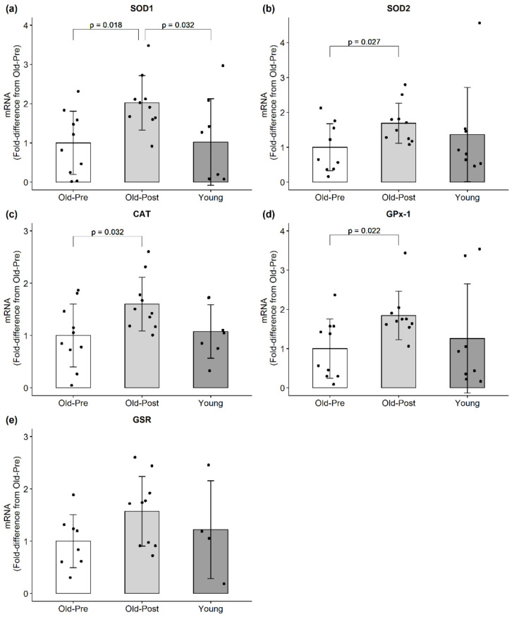
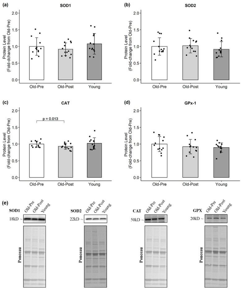
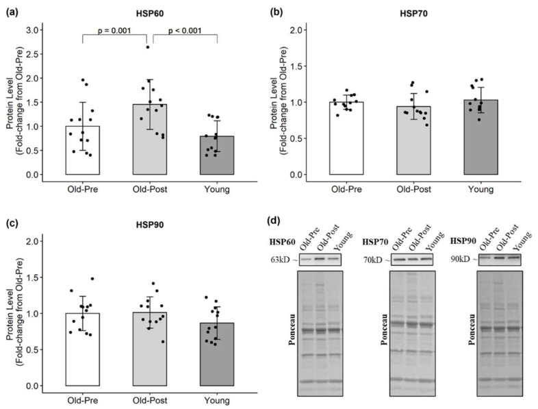
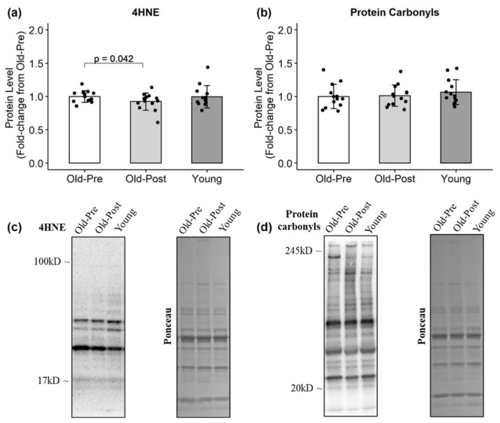
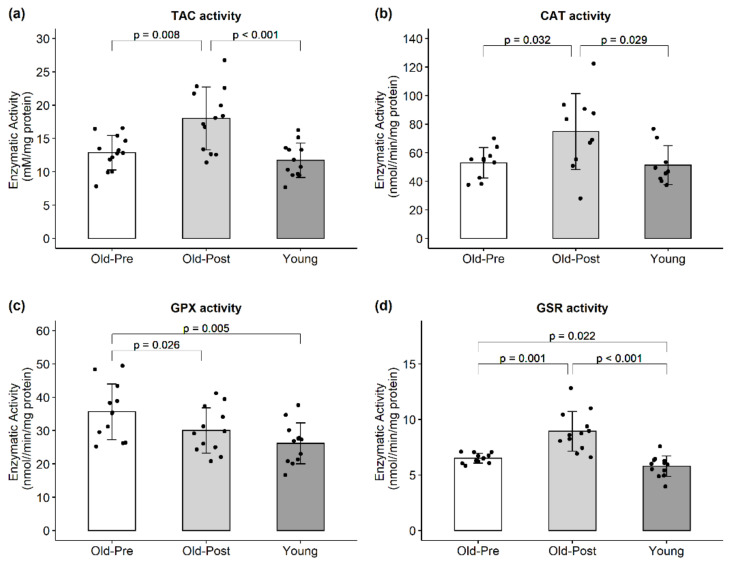
The aim of this study was to investigate the effects of resistance training (RT) on the redox status of skeletal muscle in older adults. Thirteen males aged 64 ± 9 years performed full-body RT 2x/week for 6 weeks. Muscle biopsies were obtained from the vastus lateralis prior to and following RT. The mRNA, protein, and enzymatic activity levels of various endogenous antioxidants were determined. In addition, skeletal muscle 4-hydroxynonenal and protein carbonyls were determined as markers of oxidative damage. Protein levels of heat shock proteins (HSPs) were also quantified. RT increased mRNA levels of all assayed antioxidant genes, albeit protein levels either did not change or decreased. RT increased total antioxidant capacity, catalase, and glutathione reductase activities, and decreased glutathione peroxidase activity. Lipid peroxidation also decreased and HSP60 protein increased following RT. In summary, 6 weeks of RT decreased oxidative damage and increased antioxidant enzyme activities. Our results suggest the older adult responses to RT involve multi-level (transcriptional, post-transcriptional, and post-translational) control of the redox status of skeletal muscle.

Keywords: antioxidants; exercise; oxidative damage; oxidative stress; redox homeostasis.


Conflict of interest statement

M.D.R. and K.C.Y. perform contracted studies for nutritional supplement companies, but neither they nor any of the other authors have financial or other conflicts of interest to report regarding these data. The funders had no role in the design of the study; in the collection, analyses, or interpretation of data; in the writing of the manuscript, or in the decision to publish the results.

Figures

Figure 1
Figure 1
mRNA expression of endogenous antioxidants. All data are presented as mean ± SD values, and individual data points representing each participant is superimposed on each bar graph. (a) SOD1; (b) SOD2; (c) CAT; (d) GPx-1; (e) GSR.
Figure 2
Figure 2
Protein levels of endogenous antioxidants. All data are presented as mean ± SD values, and individual data points representing each participant are superimposed on each bar graph. (a) SOD1; (b) SOD2; (c) CAT; (d) GPx-1; (e) representative Western blots.
Figure 3
Figure 3
Protein levels of HSPs. All data are presented as mean ± SD values, and individual data points representing each participant are superimposed on each bar graph. (a) HSP60; (b) HSP70; (c) HSP90; (d) representative Western blots.
Figure 4
Figure 4
Protein levels of oxidative damage markers. All data are presented as mean ± SD values, and individual data points representing each participant are superimposed on each bar graph. (a) 4HNE; (b) protein carbonyls; (c,d) representative Western blots.
Figure 5
Figure 5
Enzymatic activity of endogenous antioxidants. All data are presented as mean ± SD values, and individual data points representing each participant are superimposed on each bar graph. (a) Total antioxidant capacity; (b) CAT activity; (c) GPX activity; (d) GSR activity.

References

    1. Berger M.J., Doherty T.J. Sarcopenia: Prevalence, mechanisms, and functional consequences. Interdiscip. Top. Gerontol. 2010;37:94–114. doi: 10.1159/000319997. - DOI - PubMed
    1. Niccoli T., Partridge L. Ageing as a risk factor for disease. Curr. Biol. 2012;22:R741–R752. doi: 10.1016/j.cub.2012.07.024. - DOI - PubMed
    1. López-Otín C., Blasco M.A., Partridge L., Serrano M., Kroemer G. The hallmarks of aging. Cell. 2013;153:1194–1217. doi: 10.1016/j.cell.2013.05.039. - DOI - PMC - PubMed
    1. Dai D.-F., Chiao Y., Marcinek D.J., Szeto H.H., Rabinovitch P.S. Mitochondrial oxidative stress in aging and healthspan. Longev. Healthspan. 2014;3:6. doi: 10.1186/2046-2395-3-6. - DOI - PMC - PubMed
    1. Harman D. Aging: A theory based on free radical and radiation chemistry. J. Gerontol. 1956;11:298–300. doi: 10.1093/geronj/11.3.298. - DOI - PubMed

Grants and funding

LinkOut - more resources