. 2021 Mar 2;13(1):54.
doi: 10.1186/s13195-021-00789-5.

Association of CSF proteins with tau and amyloid β levels in asymptomatic 70-year-olds

Affiliations

Association of CSF proteins with tau and amyloid β levels in asymptomatic 70-year-olds

Julia Remnestål et al. Alzheimers Res Ther. .

Abstract

Background: Increased knowledge of the evolution of molecular changes in neurodegenerative disorders such as Alzheimer's disease (AD) is important for the understanding of disease pathophysiology and also crucial to be able to identify and validate disease biomarkers. While several biological changes that occur early in the disease development have already been recognized, the need for further characterization of the pathophysiological mechanisms behind AD still remains.

Methods: In this study, we investigated cerebrospinal fluid (CSF) levels of 104 proteins in 307 asymptomatic 70-year-olds from the H70 Gothenburg Birth Cohort Studies using a multiplexed antibody- and bead-based technology.

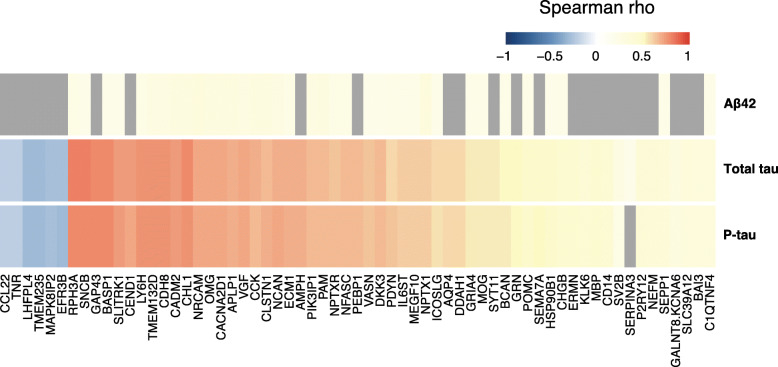
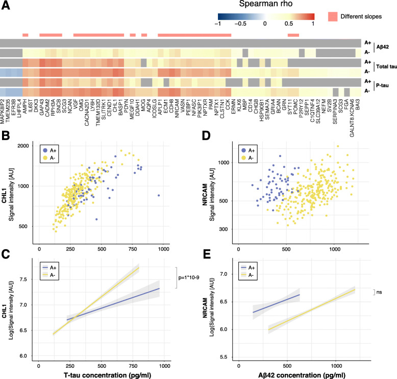
Results: The protein levels were first correlated with the core AD CSF biomarker concentrations of total tau, phospho-tau and amyloid beta (Aβ42) in all individuals. Sixty-three proteins showed significant correlations to either total tau, phospho-tau or Aβ42. Thereafter, individuals were divided based on CSF Aβ42/Aβ40 ratio and Clinical Dementia Rating (CDR) score to determine if early changes in pathology and cognition had an effect on the correlations. We compared the associations of the analysed proteins with CSF markers between groups and found 33 proteins displaying significantly different associations for amyloid-positive individuals and amyloid-negative individuals, as defined by the CSF Aβ42/Aβ40 ratio. No differences in the associations could be seen for individuals divided by CDR score.

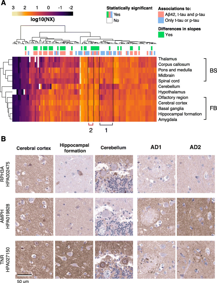
Conclusions: We identified a series of transmembrane proteins, proteins associated with or anchored to the plasma membrane, and proteins involved in or connected to synaptic vesicle transport to be associated with CSF biomarkers of amyloid and tau pathology in AD. Further studies are needed to explore these proteins' role in AD pathophysiology.

Keywords: AD pathophysiology; Affinity proteomics; Brain-enriched proteins; CSF markers; Multidisciplinary epidemiological studies; Preclinical Alzheimer’s disease.


Conflict of interest statement

HZ has served at scientific advisory boards for Denali, Roche Diagnostics, Wave, Samumed and CogRx, has given lectures in symposia sponsored by Fujirebio, Alzecure and Biogen, and is a co-founder of Brain Biomarker Solutions in Gothenburg AB, a GU Ventures-based platform company at the University of Gothenburg. KB has served as a consultant or at advisory boards for Abcam, Axon, Biogen, Lilly, MagQu, Novartis and Roche Diagnostics and is a co-founder of Brain Biomarker Solutions in Gothenburg AB (BBS), which is a part of the GU Ventures Incubator Program.

Figures

Fig. 1
Fig. 1
Protein correlation heatmap. A heatmap of all significant correlation coefficients for Aβ42, t-tau and p-tau based on protein levels from all individuals. Spearman rho values are indicated by the colour key. Non-significant correlations are presented in grey.
Fig. 2
Fig. 2
Associations with CSF markers for individuals divided by CSF Aβ42/Aβ40 ratio. a Heatmap of all significant correlation coefficients for individuals defined as A+ and A−. Spearman rho values are indicated by the colour key and grey colour represents non-significant correlations. Significant differences in slopes for association to t-tau and p-tau are indicated in pink. b Scatterplot of CHL1 levels and t-tau concentration. Both A+ and A− individuals display significant associations between CHL1 levels and t-tau concentration (A+: Spearman rho = 0.69; p = 5E−07, A−: Spearman rho = 0.80; p = 5E−56). c Linear regression revealed a significant difference between the slopes of CSF A+ and CSF A− individuals for the association between CHL1 and t-tau concentration (t = 7.13; p = 1E−09). d Scatterplot of NRCAM levels and Aβ42 concentration. A− individuals displayed a significant correlation between NRCAM levels and Aβ42 concentration (Spearman rho = 0.56, p = 4E−20) but not A+ individuals (Spearman rho = 0.36, p = 0.8). e Linear regression showed no significant difference between slopes of A+ and A− individuals for the association between NRCAM and Aβ42 concentration
Fig. 3
Fig. 3
NEFM and GAP43 levels after stratification based on CSF Aβ42/Aβ40, NfL concentration and APOE ε4 carrier status. a Visualization of NEFM levels in individuals divided by NfL concentration and CSF Aβ42/Aβ40 ratio. b Visualization of GAP43 levels in individuals divided by NfL concentration and CSF Aβ42/Aβ40 ratio. Higher levels of GAP43 were identified in Nf+A+ individuals. c Visualization of NEFM levels in individuals divided by APOE ε4 carrier status and CSF Aβ42/Aβ40 ratio. APOE ε4 carriers displayed higher levels of NEFM. d Visualization of GAP43 levels in individuals divided by APOE ε4 carrier status and CSF Aβ42/Aβ40 ratio. A+ individuals displayed higher levels of GAP43 although the trend was not statistically significant
Fig. 4
Fig. 4
Regional brain expression and tissue expression of representative proteins. a Clustering of RNA expression data showed brain region clustering of the forebrain and brainstem. Two clusters identified visually included genes with lower expression in the cerebellum compared to other brain regions and higher expression in brainstem regions compared to forebrain (Cluster 1). A few genes with lower expression in white matter-rich regions such and thalamus could also be identified (Cluster 2). b Distribution of RPH3A, AMPH and TNR in cerebral cortex, hippocampal formation and cerebellum from a healthy donor, as well as temporal cortex from two AD patients. The three selected proteins displayed a similar staining pattern in cerebral cortex as well as a general neuropil positivity in the AD tissue. Furthermore, TNR showed positive staining around the amyloid plaques

Similar articles

Cited by

References

    1. Jack CR, Jr, Knopman DS, Jagust WJ, Petersen RC, Weiner MW, Aisen PS, et al. Update on hypothetical model of Alzheimer’s disease biomarkers. Lancet Neurol. 2013;12(2):207–216. doi: 10.1016/S1474-4422(12)70291-0. - DOI - PMC - PubMed
    1. Remnestål J, Just D, Mitsios N, Fredolini C, Mulder J, Schwenk JM, et al. CSF profiling of the human brain enriched proteome reveals associations of neuromodulin and neurogranin to Alzheimer’s disease. Proteomics Clin Appl. 2016;10(12):1242–1253. doi: 10.1002/prca.201500150. - DOI - PMC - PubMed
    1. Faura J, Bustamante A, Penalba A, Giralt D, Simats A, Martinez-Saez E, et al. CCL23: a chemokine associated with progression from mild cognitive impairment to Alzheimer’s disease. J Alzheimers Dis. 2020;73(4):1585–1595. doi: 10.3233/JAD-190753. - DOI - PMC - PubMed
    1. Khoonsari PE, Shevchenko G, Herman S, Remnestal J, Giedraitis V, Brundin R, et al. Improved differential diagnosis of Alzheimer’s disease by integrating ELISA and mass spectrometry-based cerebrospinal fluid biomarkers. J Alzheimers Dis. 2019;67(2):639–651. doi: 10.3233/JAD-180855. - DOI - PMC - PubMed
    1. Whelan CD, Mattsson N, Nagle MW, Vijayaraghavan S, Hyde C, Janelidze S, et al. Multiplex proteomics identifies novel CSF and plasma biomarkers of early Alzheimer’s disease. Acta Neuropathol Commun. 2019;7(1):1–14. doi: 10.1186/s40478-019-0795-2. - DOI - PMC - PubMed

Publication types