. 2021 Mar 2;11(1):4976.
doi: 10.1038/s41598-021-83771-3.

MAPRE2 mutations result in altered human cranial neural crest migration, underlying craniofacial malformations in CSC-KT syndrome

Affiliations

MAPRE2 mutations result in altered human cranial neural crest migration, underlying craniofacial malformations in CSC-KT syndrome

Cedric Thues et al. Sci Rep. .

Abstract

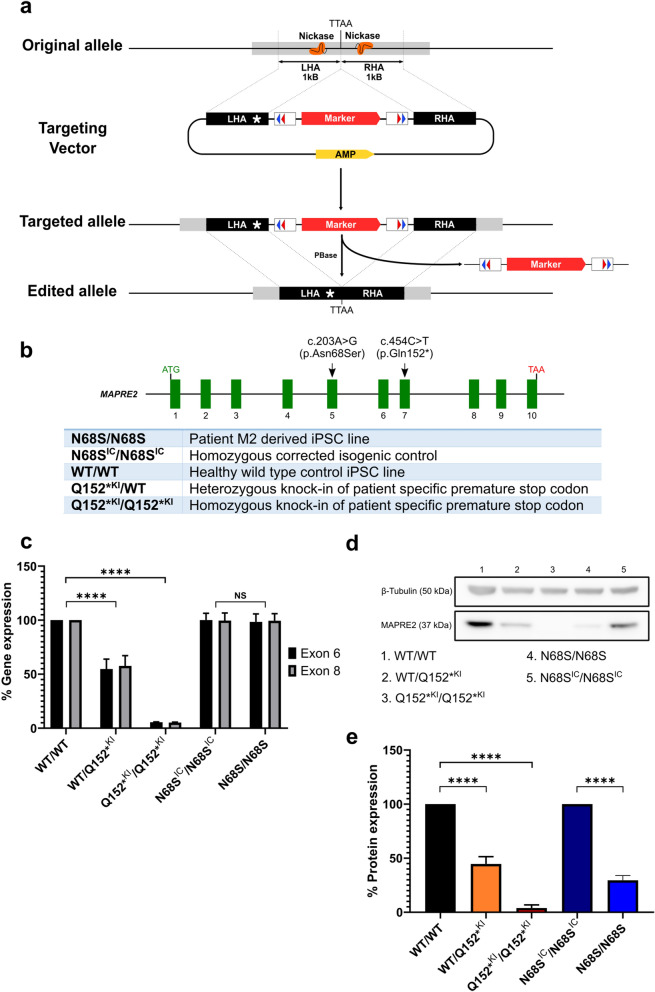
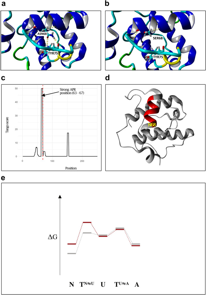
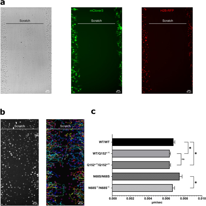
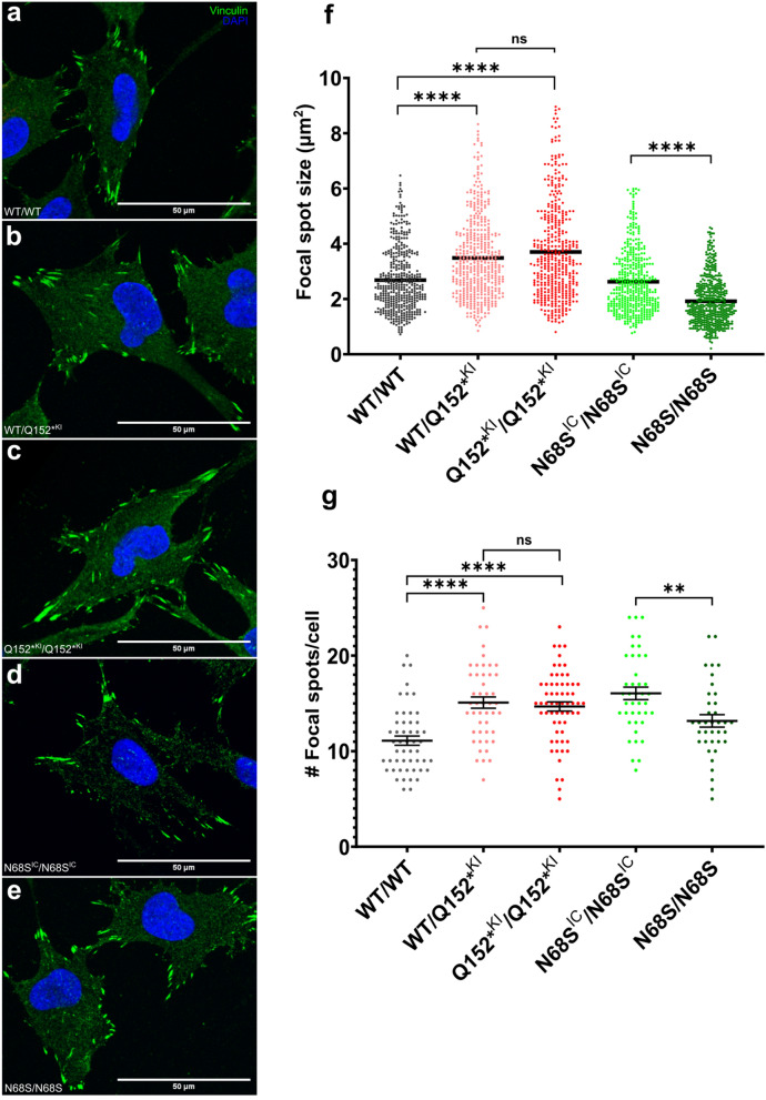
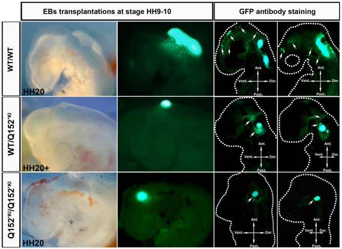
Circumferential skin creases (CSC-KT) is a rare polymalformative syndrome characterised by intellectual disability associated with skin creases on the limbs, and very characteristic craniofacial malformations. Previously, heterozygous and homozygous mutations in MAPRE2 were found to be causal for this disease. MAPRE2 encodes for a member of evolutionary conserved microtubule plus end tracking proteins, the end binding (EB) family. Unlike MAPRE1 and MAPRE3, MAPRE2 is not required for the persistent growth and stabilization of microtubules, but plays a role in other cellular processes such as mitotic progression and regulation of cell adhesion. The mutations identified in MAPRE2 all reside within the calponin homology domain, responsible to track and interact with the plus-end tip of growing microtubules, and previous data showed that altered dosage of MAPRE2 resulted in abnormal branchial arch patterning in zebrafish. In this study, we developed patient derived induced pluripotent stem cell lines for MAPRE2, together with isogenic controls, using CRISPR/Cas9 technology, and differentiated them towards neural crest cells with cranial identity. We show that changes in MAPRE2 lead to alterations in neural crest migration in vitro but also in vivo, following xenotransplantation of neural crest progenitors into developing chicken embryos. In addition, we provide evidence that changes in focal adhesion might underlie the altered cell motility of the MAPRE2 mutant cranial neural crest cells. Our data provide evidence that MAPRE2 is involved in cellular migration of cranial neural crest and offers critical insights into the mechanism underlying the craniofacial dysmorphisms and cleft palate present in CSC-KT patients. This adds the CSC-KT disorder to the growing list of neurocristopathies.


Conflict of interest statement

The authors declare no competing interests.

Figures

Figure 1
Figure 1
Overview of CRISPR-Cas9 gene editing strategy and MAPRE2 expression in newly generated cell lines. (a) General overview of CRISPR/Cas9 gene editing strategy adapted from Yusa et al.. (b) Overview of the different MAPRE2 genotypes generated. (c) RT-qPCR mRNA expression levels of MAPRE2 exon 6 and exon 8 normalized to GAPDH in non-differentiated iPSCs (n = 3). (d) Immunoblotting from non-differentiated iPSC whole cell protein lysate against MAPRE2 and β-Tubulin. Full length blot is included in Supplementary Fig. S3, f. (e) Western blot MAPRE2 protein expression levels normalized to β-Tubulin in non-differentiated iPSCs (n = 5). Q152*KI mutants are normalized to WT/WT and N68S/N68S is normalized to N68SIC/N68SIC.
Figure 2
Figure 2
Modelling of the MAPRE2 N68S mutation. (a, b) The size and hydrophobicity difference between (a) wild type (Asn) and (b) mutant (Ser) residue makes the mutant unable to form the same hydrogen bonds. (c) Aggregation propensity prediction TANGO scores per residue for wild type MAPRE2. Vertical red line indicates the position of N68. (d) Model structure of MAPRE2 with the strong APR colored in red. (e) Energetic landscape of the mechanism proposed. N68S mutation is indicated in red and wild type energy landscape in grey. N68S thermodynamically destabilizes and kinetically slows down the folding reaction, resulting in higher energies for N and TN⇋U and less natively folded protein (N native fold, U unfolded state, A aggregated state, T transition state).
Figure 3
Figure 3
In vitro migration of iPSC derived wild type and mutant cranial neural crest cells. (a) Representative images of scratched area for BJ1 wild type derived CNCC to migrate into at 0 h. Cells have been transfected with mRNA 24 h prior to imaging. From left to right: Bright field, cytosolic free-floating mClover3 and nuclei labelled with H2B-mCherry. Scale bar is 50 μm. Also see Additional Movie Files 1, 2 and 3. (b) Left: image of the same scratched area in (a) after 24 h of migration visualizing labeled nuclei with H2B-mCherry. Right: Representative image of tracking the migration path of individual neural crest cells using ImageJ plugin TrackMate. (c) Quantification of the neural crest migration speed. Cells carrying either the heterozygous or the homozygous Q152* mutation (Q152*KI/WT, Q152*KI/Q152*KI) displayed a lower migration speed compared to their isogenic control line WT/WT. No difference between the full knock down Q152*KI/Q152*KI of MAPRE2 compared to the heterozygous knock down Q152*KI/WT was noted (One-Way ANOVA, Tukey test, P = 0.045, P = 0.0423 and P = 0.9996, respectively). In contrast, the N68S/N68S mutant NC cells migrated faster than their respective isogenic control line N68SIC/N68SIC (Unpaired t-test, P = 0.044). N = 11 for each genotype.
Figure 4
Figure 4
Focal adhesion in iPSC derived cranial neural crest. (ae) Immunofluorescence staining for DAPI (blue) and focal adhesion marker vinculin (green) in iPSC derived CNCC plated into fibronectin coated culture plates. Cells were imaged using confocal microscopy. Scale bar is 50 μm. (f) Quantification and size distribution of FA spots. N > 400 per genotype. An increased labelling of focal adhesion spots in both Q152*KI/WT and Q152*KI/Q152*KI mutant lines is observed compared to their isogenic control WT/WT with no difference between Q152*KI/WT and Q152*KI/Q152*KI (Kruskal–Wallis test, P < 0.0001, P < 0.0001, P > 0.9999, respectively). Less labelling of focal adhesion spots and a decrease in spot size for the N68S/N68S mutant is observed compared to its isogenic control N68SIC/N68SIC (Mann–Whitney test; P < 0.0001). (g) Quantification of number of FA spots per cell. N =  > 37 per genotype. An increase in focal adhesion spots in both Q152*KI/WT and Q152*KI/Q152*KI mutant lines is observed compared to their isogenic control WT/WT with no difference between Q152*KI/WT and Q152*KI/Q152*KI (One-way ANOVA, P < 0.0001, P < 0.0001, P > 0.9999, respectively). Fewer focal adhesion spots for the N68S/N68S mutant is observed compared to its isogenic control N68SIC/N68SIC (Unpaired t test; P = 0.0026).
Figure 5
Figure 5
Absent migration of xenotransplanted MAPRE2 deficient neural crest progenitors in chicken embryos. Representative whole mount bright field and fluorescent images of chicken embryos at development stage HH 20 with corresponding sagittal sections. Embryos have been transplanted with EBs at day 5 of neural crest differentiation, generated from EmGFP tagged MAPRE2 deficient iPSCs. Sagittal sections have been stained against endogenous EmGFP. WT/WT cells are observed migrating into the branchial arches and facial mesenchyme on whole mount and in sections, indicated by white arrows. Q152*KI/WT cells emerge from the EB but fail to migrate away towards the branchial arches and facial mesenchyme (indicated by white arrows). Complete MAPRE2 knock out cells (Q152*KI/Q152*KI) fail to emerge from the EB and migrate. (Ant. anterior, Vent. ventral, Dor. dorsal, Post. posterior).

References

    1. Isrie M, et al. Mutations in either TUBB or MAPRE2 cause circumferential skin creases kunze type. Am. J. Hum. Genet. 2015;97:790–800. doi: 10.1016/j.ajhg.2015.10.014. - DOI - PMC - PubMed
    1. Wouters L, et al. Circumferential skin creases, cleft palate, typical face, intellectual disability and growth delay: ‘Circumferential skin creases Kunze type’. Eur. J. Med. Genet. 2011;54:236–240. doi: 10.1016/j.ejmg.2011.01.003. - DOI - PubMed
    1. Su LK, Qi Y. Characterization of human MAPRE genes and their proteins. Genomics. 2001;71:142–149. doi: 10.1006/geno.2000.6428. - DOI - PubMed
    1. Galjart N. Plus-end-tracking proteins and their interactions at microtubule ends. Curr. Biol. 2010;20:R528–R537. doi: 10.1016/j.cub.2010.05.022. - DOI - PubMed
    1. Akhmanova A, Steinmetz MO. Tracking the ends: A dynamic protein network controls the fate of microtubule tips. Nat. Rev. Mol. Cell Biol. 2008;9:309–322. doi: 10.1038/nrm2369. - DOI - PubMed

Publication types

Substances