Review
. 2021 Mar 2;5(1):15.
doi: 10.1038/s41698-021-00155-6.

Structural variant detection in cancer genomes: computational challenges and perspectives for precision oncology

Affiliations
Review

Structural variant detection in cancer genomes: computational challenges and perspectives for precision oncology

Ianthe A E M van Belzen et al. NPJ Precis Oncol. .

Abstract

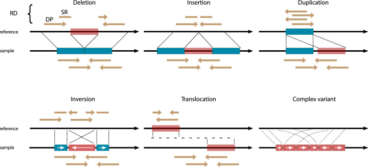
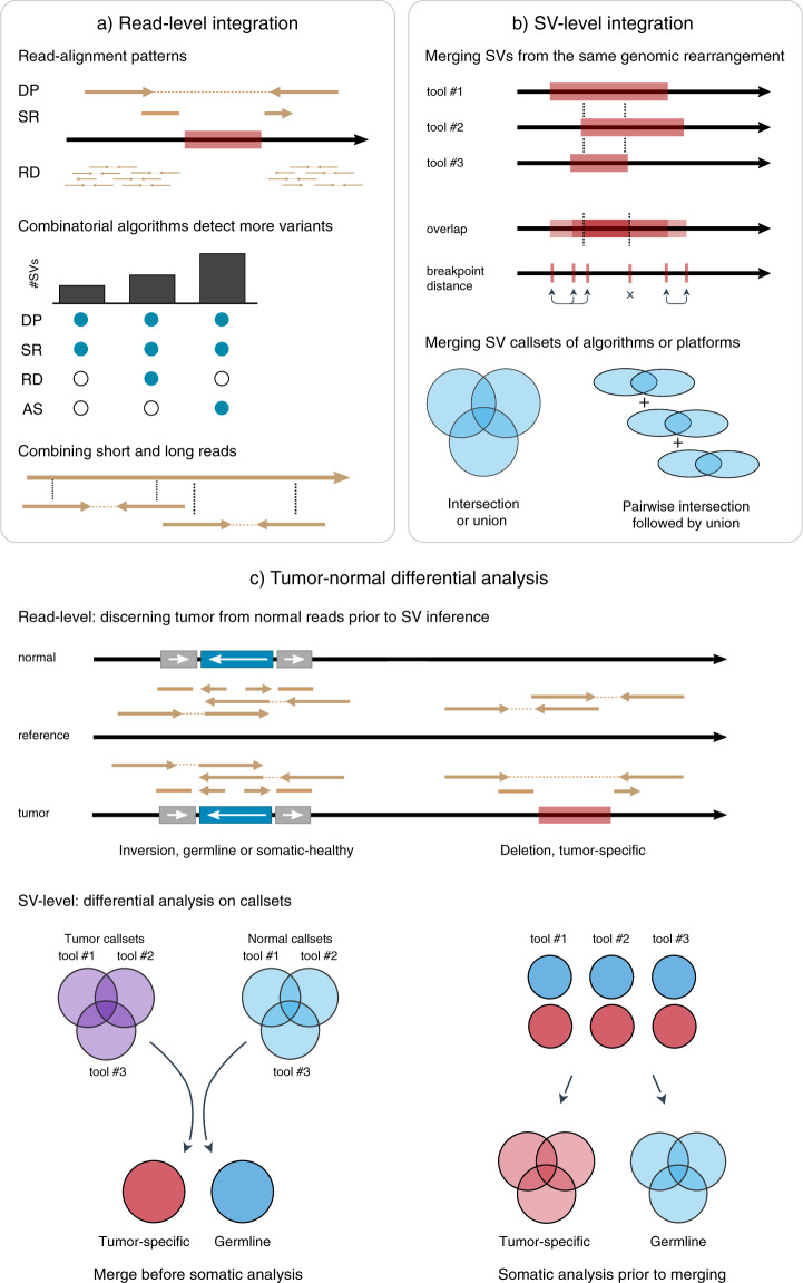
Cancer is generally characterized by acquired genomic aberrations in a broad spectrum of types and sizes, ranging from single nucleotide variants to structural variants (SVs). At least 30% of cancers have a known pathogenic SV used in diagnosis or treatment stratification. However, research into the role of SVs in cancer has been limited due to difficulties in detection. Biological and computational challenges confound SV detection in cancer samples, including intratumor heterogeneity, polyploidy, and distinguishing tumor-specific SVs from germline and somatic variants present in healthy cells. Classification of tumor-specific SVs is challenging due to inconsistencies in detected breakpoints, derived variant types and biological complexity of some rearrangements. Full-spectrum SV detection with high recall and precision requires integration of multiple algorithms and sequencing technologies to rescue variants that are difficult to resolve through individual methods. Here, we explore current strategies for integrating SV callsets and to enable the use of tumor-specific SVs in precision oncology.


Conflict of interest statement

The authors declare no competing interests.

Figures

Fig. 1
Fig. 1. Major SV types and their characteristic read-alignment patterns.
Alignment of paired-end sequencing reads to a reference genome is used to infer sites of discontinuity or breakpoints. Structural variants (SVs) are generally defined as larger than 50 base pairs and further classified in five major SV types: deletions, insertions of non-reference sequence or mobile elements, duplications, inversions and translocations. Clusters of breakpoints in a genomic region which cannot be classified are considered “complex SVs” and likely result from either progressive rearrangements or a major genomic disturbance. SVs (red blocks) are characterized by patterns in breakpoints and reads aligned to flanking reference sequences (blue blocks). The reads directly below the sample DNA strand represent the distance and orientation at which they are generated during sequencing. If the reads align differently than expected to the reference strand this is indicative of an SV. Changes in read depth (RD) or coverage indicate mostly larger duplications or deletions and are useful for detecting copy number variants (CNVs). Discordant pairs (DP) align to the reference at a different relative distance or orientation than expected. DPs are best suited for detecting large SVs such as inter-chromosomal translocations or inversions. Split reads (SR) span breakpoints and can only be partially aligned. SR can detect small variants with base-pair resolution, especially those smaller than the size of the read.
Fig. 2
Fig. 2. Data integration to improve tumor-specific SV detection.
a Alignment of sequencing data against a reference is used to infer SVs by detecting aberrant patterns of read-alignment: discordant pairs (DP), split reads (SR), read depth (RD) and (local) assembly (top, see also Fig. 1). Algorithms that combine multiple read-alignment patterns can resolve more SVs (middle). Likewise, read-level integration of technologies can aid SV detection, i.e., combining short and long reads (bottom). b Comparison of SV callsets requires merging variants from the same genomic rearrangement based on e.g., reciprocal overlap or breakpoint distance (top). These merging approaches can yield different outcomes as shown by how only a small segment of the deletion overlaps between tools and not all breakpoints could be matched. Intersection of callsets identifies the SVs with support from multiple algorithms or technologies. Alternatively, sensitivity can be increased by taking the union of callsets or their pairwise intersections (bottom). c Identification of tumor-specific SVs (red) requires tumor-normal differential analysis of reads or events. A tumor sample (purple) is expected to contain tumor-specific variants (red, bottom stand), as well as germline variants (blue, top strand). Tumor/normal reads can be distinguished prior to SV inference or afterwards by comparison of the variants or breakpoints as in b. If multiple SV tools are used, differential analysis can be done after merging tumor and normal callsets (bottom left) or first by using each algorithm’s somatic filtering feature (bottom right).

References

    1. Vogelstein B, Kinzler KW. Cancer genes and the pathways they control. Nat. Med. 2004;10:789–799. doi: 10.1038/nm1087. - DOI - PubMed
    1. Aplan PD. Causes of oncogenic chromosomal translocation. Trends Genet. 2006;22:46–55. doi: 10.1016/j.tig.2005.10.002. - DOI - PMC - PubMed
    1. Sudmant PH, et al. An integrated map of structural variation in 2,504 human genomes. Nature. 2015;526:75. doi: 10.1038/nature15394. - DOI - PMC - PubMed
    1. Ho SS, Urban AE, Mills RE. Structural variation in the sequencing era. Nat. Rev. Genet. 2019;21:1–19. - PMC - PubMed
    1. Calabrese C, et al. Genomic basis for RNA alterations in cancer. Nature. 2020;578:129–136. doi: 10.1038/s41586-020-1970-0. - DOI - PMC - PubMed