Review
. 2021 Feb 4:9:tkaa047.
doi: 10.1093/burnst/tkaa047. eCollection 2021 Jan.

The pathogenesis and diagnosis of sepsis post burn injury

Affiliations
Review

The pathogenesis and diagnosis of sepsis post burn injury

Pengju Zhang et al. Burns Trauma. .

Abstract

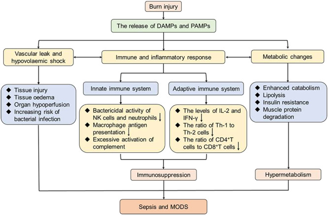
Burn is an under-appreciated trauma that is associated with unacceptably high morbidity and mortality. Although the survival rate after devastating burn injuries has continued to increase in previous decades due to medical advances in burn wound care, nutritional and fluid resuscitation and improved infection control practices, there are still large numbers of patients at a high risk of death. One of the most common complications of burn is sepsis, which is defined as "severe organ dysfunction attributed to host's disordered response to infection" and is the primary cause of death in burn patients. Indeed, burn injuries are accompanied by a series of events that lead to sepsis and multiple organ dysfunction syndrome, such as a hypovolaemic state, immune and inflammatory responses and metabolic changes. Therefore, clear diagnostic criteria and predictive biomarkers are especially important in the prevention and treatment of sepsis and septic shock. In this review, we focus on the pathogenesis of burn wound infection and the post-burn events leading to sepsis. Moreover, the clinical and promising biomarkers of burn sepsis will also be summarized.

Keywords: Biomaker; Burn; Hypermetabolism; Immune dysregulation; Infection; Inflammation; Multiple organ dysfunction syndrome; Sepsis; Septic shock; Trauma.


Figures

Figure 1.
Figure 1.
Zones of burn injury for different depths. First-degree burns only involve the epidermis (the uppermost layer of the skin); the skin becomes red and painful, but this is limited in duration. Burns that affect the dermis (the underlying skin layer) are classed as partial-thickness burns, which are frequently accompanied by the formation of painful blisters that increase the risk of infection. Partial-thickness burns can be divided into superficial partial-thickness burns, which are painful, moist, hyperemic and blanch, and deep partial-thickness burns which are less sensate, drier and do not blanch. Full-thickness burns extend through the full dermis and require surgical management due to high risk of infection. Burns extending into deeper tissues (such as muscle or even bone) are defined as fourth-degree burns and are usually blackened and often result in loss of the burned tissues
Figure 2.
Figure 2.
Lund and Browder diagrams for estimation of total burned surface area (TBSA). The “rule of nines” (using multiples of 9) is frequently used to assess the proportion of TBSA affected in adults and to help guide immediate treatment decisions based on burn size. However, the rule of nines is inaccurate in children due to different head-to-body size ratios at different ages. Lund and Browder diagrams are therefore more suitable for assessing of the proportion of TBSA affected in both children and adults. The body areas are separated into different regions (including anterior and posterior) by dashed lines and the numbers are percentages of the TBSA. For instance, 19 in the diagram for children aged 1–4 years relates to the face, neck and head that make up 19% of theTBSA
Figure 3.
Figure 3.
A series of pathogenic events responsible for sepsis post burn injury. Following severe burn injury, damaged tissues lead to the release of endogenous DAMPs (such as double-stranded RNA and mitochondrial DNA) and exogenous PAMPs (such as lipopolysaccharides and peptidoglycans). Subsequently, PAMPs can result in vascular leak and hypovolemic shock, immune and inflammatory responses and metabolic changes. Vascular leak causes tissue edema, organ hypoperfusion and increasing risk of bacterial infection. Meanwhile, the excessive inflammatory response leads to immunosuppression by inhibition of the innate and adaptive immune systems. Moreover, hypermetabolism emerges in the form of enhanced catabolism, lipolysis, insulin resistance and muscle protein degradation. These events contribute to the susceptibility of the burn patients to sepsis and MODS. DAMPs damage-associated molecular patterns, PAMPs pathogen-associated molecular pattern molecules, NK natural killer, IL-2 interleukin 2, IFN-γ interferon γ, Th-1 helper T lymphocyte 1, Th-2 helper T lymphocyte 2, MODS multiple organ dysfunction syndrome
Figure 4.
Figure 4.
The host response to infection and during sepsis. After infection, PAMPs are released and interact with cell-surface, intracellular and even secreted PRRs, including toll-like receptors, nucleotide-binding oligomerization domain-like receptors, retinoic acid-inducible gene-like receptors and C-type lectin receptors. The interaction between PAMPs and PRRs can result in cytokine secretion, immune cell apoptosis and the activation of the complement system. In some burn patients, these events lead to the simultaneous imbalanced activation of proinflammatory response (excessive inflammation) and anti-inflammatory response (immune suppression). Excessive inflammation can result in the dysfunction of the endothelial barrier, microvascular thrombi and further injuries. Immunosuppression causes decreased bactericidal activity of neutrophils and NK cells, decreased phagocytosis and antigen presentation of macrophages and impaired innate immune system response. Taken together, excessive inflammation and immunosuppression contribute to a greatly increased risk for sepsis and organ dysfunction. PAMPs pathogen-associated molecular pattern molecules, PRR pattern recognition receptors, DAMPs damage-associated molecular patterns, NK natural killer, Th-2 helper T lymphocyte 2

References

    1. Jeschke MG, Baar ME, Choudhry MA, Chung KK, Gibran NS, Logsetty S. Burn injury. Nat. Rev. Dis. Primers. 2020. 10.1038/s41572-020-0145-5. - DOI - PMC - PubMed
    1. National Burn Repository 2019 Update , Report of data from 2009–2018 ameriburn.site- ym.com [Internet]. 2019. Available from: https://ameriburn.site-ym.com/store/ViewProduct.aspx?id=14191872.
    1. Pereira RF, Barrias CC, Granja PL, Bartolo PJ. Advanced biofabrication strategies for skin regeneration and repair. Nanomedicine (Lond.) 2013;8:603–21. - PubMed
    1. Peck MD. Epidemiology of burns throughout the world. Part I: distribution and risk factors. Burns 2011;37:1087–100. - PubMed
    1. Cioffi WG. deLemos RA, Coalson JJ, Gerstmann DA, Pruitt BA Jr. decreased pulmonary damage in primates with inhalation injury treated with high-frequency ventilation. Ann. Surg. 1993;218:328–35discussion 35-7. - PMC - PubMed