. 2020 Feb 20;10(4):e3529.
doi: 10.21769/BioProtoc.3529.

Hippocampal Unicellular Recordings and Hippocampal-dependent Innate Behaviors in an Adolescent Mouse Model of Alzheimer's disease

Affiliations

Hippocampal Unicellular Recordings and Hippocampal-dependent Innate Behaviors in an Adolescent Mouse Model of Alzheimer's disease

Siddhartha Mondragón-Rodríguez et al. Bio Protoc. .

Abstract

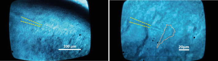
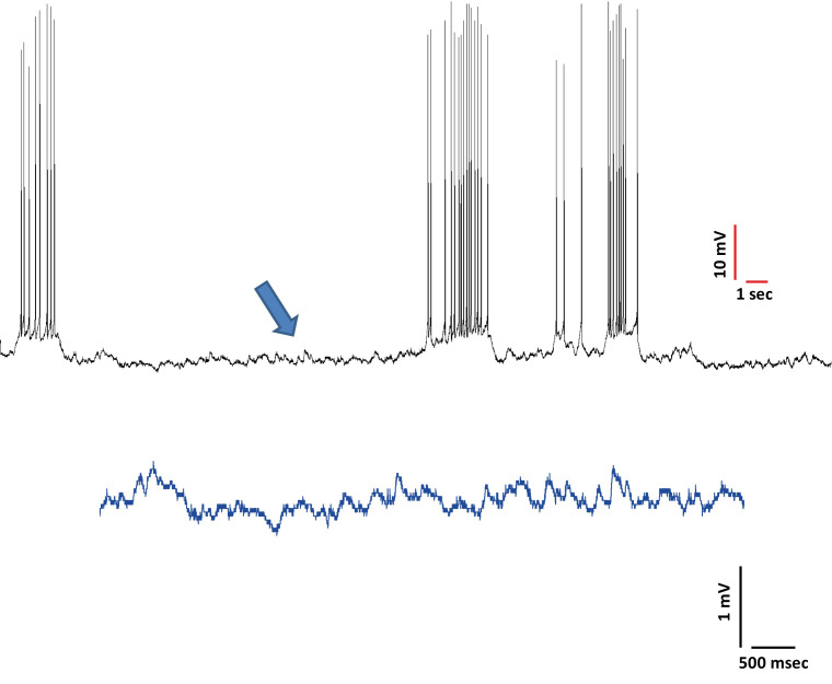
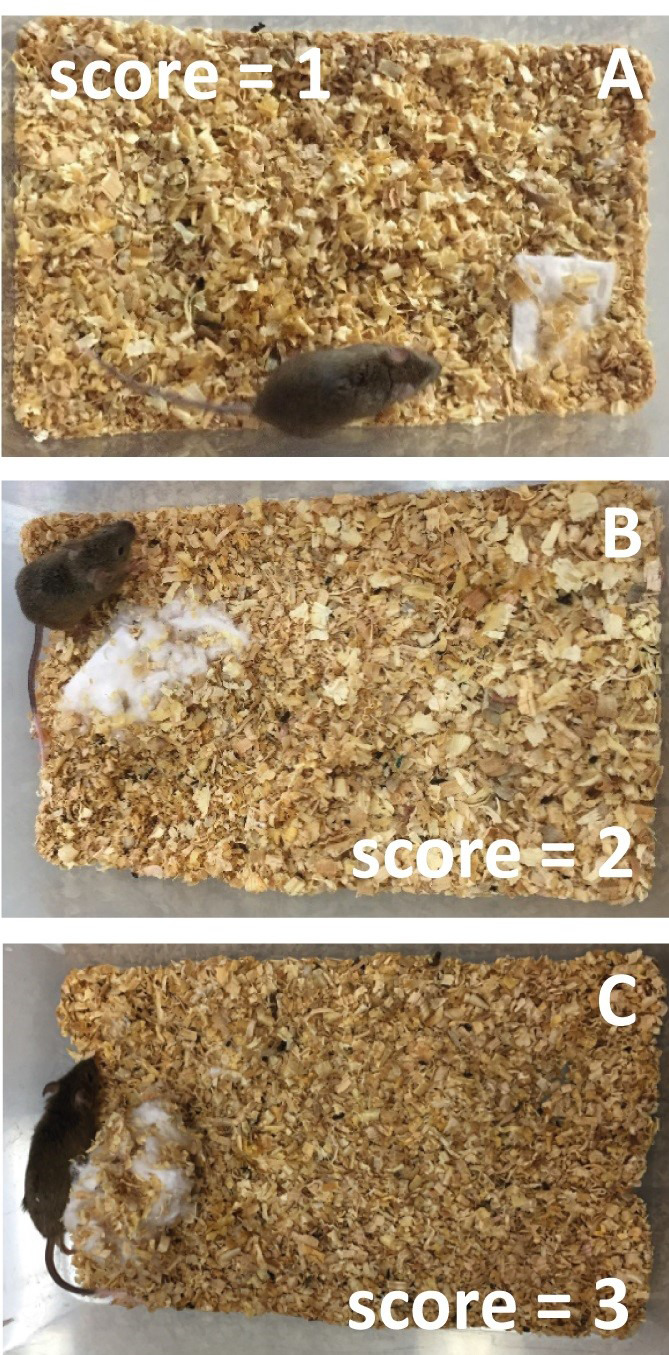
Transgenic mice have been used to make valuable contributions to the field of neuroscience and model neurological diseases. The simultaneous functional analysis of hippocampal cell activity combined with hippocampal dependent innate task evaluations provides a reliable experimental approach to detect fine changes during early phases of neurodegeneration. To this aim, we used a merge of patch-clamp with two hippocampal innate behavior tasks. With this experimental approach, whole-cell recordings of CA1 pyramidal cells, combined with hippocampal-dependent innate behaviors, have been crucial for evaluating the early mechanism of neurodegeneration and its consequences. Here, we present our protocol for ex vivo whole-cell recordings of CA1 pyramidal cells and hippocampal dependent innate behaviors in an adolescent (p30) mice.

Keywords: Hippocampus; Innate behavior; Neurodegeneration; Patch-clamp; Pyramidal cells; Subthreshold oscillations and Alzheimer’s disease.


Conflict of interest statement

Competing interestsThe authors declare no competing financial interests.

Figures

Figure 1.
Figure 1.. Homemade plastic black tube used as a burrow
Figure 2.
Figure 2.. Place an animal in an individual cage with wood-chip bedding following by a cotton square 60 min later
(scale bar = 5 cm)
Figure 3.
Figure 3.. Plastic black tube with pellets displaced at time 0, 8 a.m. and 9 p.m.
Figure 4.
Figure 4.. To obtain para-sagittal slices (see sagittal cut, red dotted line), separate the brain hemispheres and glue them by their medial portion at a 30° angle.
First, glue the brain to a piece of filter paper and then, glue the filter paper (with the brain) to the agar block (see sagittal slice sequence). The agar block is glue to the adaptor tray (R = rostral, D = dorsal, C = caudal, V = ventral).
Figure 5.
Figure 5.. Sagittal slice sequence.
With the razor blade, separate the brain hemispheres (A), glue the brain to a piece of filter paper (B) and then, glue the filter paper (with the brain) to the agar block (C to E). Finally, transfer the plate into the slicing chamber of the vibratome and lower the blade holder (F). Once the slice is freed, transfer it to the nest beaker with recovery aCSF.
Figure 6.
Figure 6.. Incubate sagittal slices in aCSF bubbled with carbogen at room temperature for at least one hour for recovery.
The nest beaker could be prepared according to Papouin et al., 2018 .
Figure 7.
Figure 7.. Electrophysiology whole-cell patch-clamp set up and microscope
Figure 8.
Figure 8.. Slice holder and recording chamber.
The slice holder is made of a bent and flattened stainless-steel wire crossed by horizontal nylon fibers (around 3 mm apart). The recording chamber is made of acrylic (measuring length in centimeters, cm).
Figure 9.
Figure 9.. Mounting and holding the brain slice containing the hippocampus onto the recording chamber.
The brain slice containing the hippocampal formation is placed onto the recording chamber and immobilized with the holder, leaving the recording area free for electrode location but fixed to maintain stable recording while the slice is perfused at high speed (17-20 ml/min).
Figure 10.
Figure 10.. Images of the CA1 pyramidal cell layer visualized through a differential interference contrast (DIC) microscope.
The visual patch-clamp technique, in the whole-cell configuration, is achieved with pipette electrodes (highlighted with the dotted line) located on the cell’s soma (10x and 40x magnification on the left and right, respectively).
Figure 11.
Figure 11.. Characterization of the evoked firing pattern of a recorded whole-cell neuron.
Injecting a one-second, 30 pA depolarizing current pulse (lower trace) induced a train of action potentials (upper trace), which exhibit the adaptation phenomenon (reduction in firing frequency during steady stimulation).
Figure 12.
Figure 12.. Bring the membrane potential to the level just below the level where action potentials are triggered (see arrow)
Figure 13.
Figure 13.. Nestlet is not noticeably made (A, see cotton square), nestlet partially torn up (B, see cotton square) and a near perfect nest (C, see cotton square)
Figure 14.
Figure 14.. For power spectrum analysis of the membrane potential, analyze segments using a Fast Fourier Transform algorithm with a Hamming window in Clampfit

Similar articles

Cited by

References

    1. Deacon R.(2012). Assessing burrowing, nest construction, and hoarding in mice. J Vis Exp(59): e2607. - PMC - PubMed
    1. Deacon R. M., Raley J. M., Perry V. H. and Rawlins J. N.(2001). Burrowing into prion disease. Neuroreport 12(9): 2053-2057. - PubMed
    1. Gjendal K., Ottesen J. L., Olsson I. A. S. and Sørensen D. B.(2019). Burrowing and nest building activity in mice after exposure to grid floor, isoflurane or ip injections. Physiol Behav 206: 59-66. - PubMed
    1. Hashimoto S and Saido T. C.(2018). Critical review: involvement of endoplasmic reticulum stress in the aetiology of Alzheimer’s disease. Open Biol. 8(4): 180024. - PMC - PubMed
    1. Ittner L. M., Ke Y. D., Delerue F., Bi M., Gladbach A., van Eersel J., Wolfing H., Chieng B. C., Christie M. J., Napier I. A., Eckert A., Staufenbiel M., Hardeman E. and Gotz J.(2010). Dendritic function of tau mediates amyloid-beta toxicity in Alzheimer's disease mouse models. Cell 142(3): 387-397. - PubMed

LinkOut - more resources