. 2019 Jan 20;9(2):e3142.
doi: 10.21769/BioProtoc.3142.

Click Chemistry (CuAAC) and Detection of Tagged de novo Synthesized Proteins in Drosophila

Affiliations

Click Chemistry (CuAAC) and Detection of Tagged de novo Synthesized Proteins in Drosophila

Kathrin Marter et al. Bio Protoc. .

Abstract

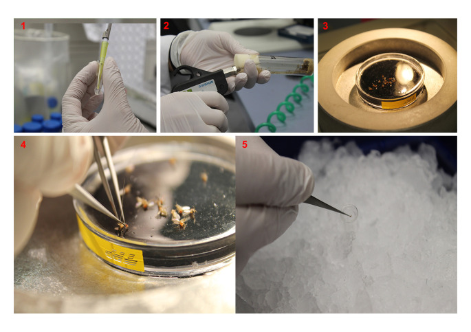
Copper-catalyzed azide-alkyne-cycloaddition (CuAAC), also known as 'click chemistry' serves as a technique for bio-orthogonal, that is, bio-compatible labeling of macromolecules including proteins or lipids. Click chemistry has been widely used to covalently, selectively, and efficiently attach probes such as fluorophores or biotin to small bio-orthogonal chemical reporter groups introduced into macromolecules. In bio-orthogonal non-canonical amino acid tagging (BONCAT) and fluorescent non-canonical amino acid tagging (FUNCAT) proteins are metabolically labeled with a non-canonical, azide-bearing amino acid and subsequently CuAAC-clicked either to an alkyne-bearing biotin (BONCAT) for protein purification, Western blot, or mass spectrometry analyses or to an alkyne-bearing fluorophore (FUNCAT) for immunohistochemistry. In combination with mass spectrometry, these kinds of labeling and tagging strategies are a suitable option to identify and characterize specific proteomes in living organisms without the need of prior cell sorting. Here, we provide detailed protocols for FUNCAT and BONCAT click chemistry and the detection of tagged de novo synthesized proteins in Drosophila melanogaster.

Keywords: Bio-orthogonal chemical reporters; Click chemistry; CuAAC; Drosophila melanogaster; Protein synthesis; Protein tagging; Proteomic profiling.


Conflict of interest statement

Competing interestsThe authors declare no conflicts of interest or competing interests.

Figures

Video 1.
Video 1.. Larval body wall dissection at 3-fold time-lapse.
A wet larva is placed on the dissection chamber shown in the supplementary figure. Using the central, straight clips the animal is immobilized and moderately stretched, dorsal side up. The animal is then covered with Ca2+-free saline. Using the spring scissors a hole is squeezed in at about 75% length, serving as a starting point for cutting along the dorsal midline, first direction mouth hook and then towards the posterior end. Short incisions are made to the right and the left at either end. To avoid scratching the body wall muscles and harming the larval brain with the blades, the scissors are slightly lifted during the cutting. Once cut, the larva is pushed a bit to squeeze out inner organs such as the gut and fat body and thus to ease their removal. The lateral clips are then used to carefully span the body wall.
Figure 1.
Figure 1.. Materials and tools for larval body wall dissection.
include Ca2+-free saline, a tap water- or saline-containing dish for rinsing larvae prior to dissection, fine forceps (e.g., Dumont #5) and spring scissors (e.g., Vannas-type, FST 15002-08). As an alternative to the widely used combination of PDMS plates with sharp tungsten needles, dissections can be carried out on a chamber (~5 x 8 cm2) that is based on a magnetic foil with a central hole of about 2 cm diameter, glued to a 1 to 2 mm thick glass slide. Stainless steel pins glued to iron-based holders and with sharp, hook-like endings are used as movable clips.
Figure 2.
Figure 2.. Cell-type-specific in situ labeling of glial proteins via FUNCAT.
ANL incorporation into larval proteins is monitored via FUNCAT on targeted expression of dMetRSL262G-EGFP in glia cells ( repo-Gal4/UAS-dMetRSL262G-EGFP ) at larval neuromuscular junctions (muscles 6/7, segment A2). Co-staining with the neuron-specific marker anti-HRP reveals that, wherever nerve terminal boutons (b) of motor neurons (m) and glial protrusions (g) are in close contact, ANL-TAMRA signal is indeed restricted to the dMetRSL262G-EGFP-expressing glial cell. Notably, dMetRSL262G-EGFP is predominantly found in the cytosol, whereas TAMRA-harboring proteins are detected throughout the glia cell including nuclei (n) and glial protrusions (g) in close proximity to the first synaptic boutons. Scale bar is 5 µm.
Video 2.
Video 2.. Fly head dissection.
First, a glass homogenizer containing 50 µl homogenization buffer is put on ice. Flies are anesthetized with CO2 in the vial for a few seconds. Once the flies are immobile, they are transferred to a PDMS dissection plate, which is placed on top of a cold plate. Using a pair of fine forceps the abdomen is hold, and the head is gently pinched off with a second pair of forceps or fine scissors. Finally, the head is directly transferred into the glass homogenizer containing homogenization buffer. Continue until a total of 20 dissected heads are collected in the homogenizer.
Figure 3.
Figure 3.. Fly head dissection.
A. Prepare a glass homogenizer containing 50 µl homogenization buffer, put it on ice. B. Anesthetize flies with CO2. C. Transfer anesthetized flies to a PDMS dissection plate, which is placed on top of a cold plate. D. Use fine forceps to keep hold of the abdomen and a second pair of forceps or fine scissor to gently pinch off/cut off the fly head. E. Transfer the head directly into the glass homogenizer containing homogenization buffer. Repeat steps (figures 3D and 3E) for a total of 20 dissected heads.
Figure 4.
Figure 4.. Western blot protein visualization and identification after chronic ANL incorporation into neuronal protein and bio-orthogonal non-canonical amino acid tagging (BONCAT).
Heads from adult flies (a) chronically exposed to ANL and (b) expressing the dMetRSL262G pan-neuronally ( elavC155-Gal4, UAS-dMetRSL262G-EGFP , see our detailed bio-protocol on cell-type specific metabolic labeling of proteins with azidonorleucine in Drosophila [ Erdmann et al., 2017 ]) were lysed (see ‘protein extraction’, Part II B), the ANL incorporated in the proteins was clicked to a biotin tag (see ‘click chemistry’, Part II C) and desalted (see ‘desalting’, Part II D). The protein concentration was normalized (see ‘adjustment of protein concentrations’, Part II E) before NeutrAvidin purification (Part II F). The representative Western blot shows the input fraction (lysate before NeutrAvidin purification), the unbound fraction (no ANL-containing proteins), and the eluate fraction (enriched ANL-labeled proteins after NeutrAvidin purification) at the global protein level (anti-biotin antibody) and for a selected candidate neuronal protein (anti-synapsin antibody). The input fraction contains all protein, that is, biotinylated and non-biotinylated protein. Therefore, the biotin signal of samples from flies exposed to ANL is diluted to approximately 1:10 depending on the respective protein compared to the same sample after NeutrAvidin purification (eluate fraction). Note that no biotin signal is observed in samples from flies not exposed to ANL, demonstrating a proper signal-to-noise ratio of the purification step. Similarly, no biotin signal is observed in the unbound fraction, demonstrating an appropriate amount of NeutrAvidinTM agarose. The relatively high synapsin signal in the unbound fraction shows that not all synapsin is biotinylated, thus that not all synapsin has ANL incorporated. This result is explained by the relatively non-invasive nature of our technique as the endogenous MetRS is not knocked out and loading methionine onto the Met-tRNA and consequently methionine into elongating polypeptide chains at the ribosome.

References

    1. Agard N. J., Prescher J. A. and Bertozzi C. R.(2004). A strain-promoted[3 + 2] azide-alkyne cycloaddition for covalent modification of biomolecules in living systems. J Am Chem Soc 126(46): 15046-15047. - PubMed
    1. Beatty K. E., Liu J. C., Xie F., Dieterich D. C., Schuman E. M., Wang Q. and Tirrell D. A.(2006). Fluorescence visualization of newly synthesized proteins in mammalian cells. Angew Chem Int Ed Engl 45(44): 7364-7367. - PubMed
    1. Brent J. R., Werner K. M. and McCabe B. D.(2009). Drosophila larval NMJ dissection . J Vis Exp(24): e1107. - PMC - PubMed
    1. Dieterich D. C.(2010). Chemical reporters for the illumination of protein and cell dynamics. Curr Opin Neurobiol 20(5): 623-630. - PubMed
    1. Dieterich D. C., Hodas J. J., Gouzer G., Shadrin I. Y., Ngo J. T., Triller A., Tirrell D. A. and Schuman E. M.(2010). In situ visualization and dynamics of newly synthesized proteins in rat hippocampal neurons . Nat Neurosci 13(7): 897-905. - PMC - PubMed

LinkOut - more resources