

Immunohistochemical Identification of Muscle Fiber Types in Mice Tibialis Anterior Sections
- PMID: 33654901
- PMCID: PMC7853988
- DOI: 10.21769/BioProtoc.3400
Immunohistochemical Identification of Muscle Fiber Types in Mice Tibialis Anterior Sections
Abstract
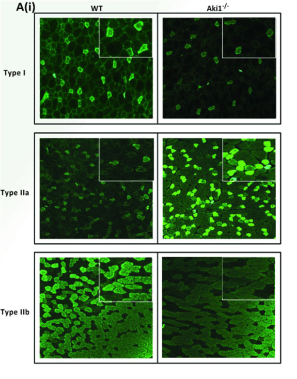
Mammalian skeletal muscle is a metabolically active tissue that is made up of different types of muscle fibers. These myofibers are made up of important contractile proteins that provide force during contraction of the muscle like actin and myosin. Murine myofibers have been classified into 4 types: Type I, Type IIa, Type IIb and Type IIX. Each muscle fiber has been identified with specific type of MyHC expressed, which in turn gives differential contractility to the muscle. There have been well-known methodologies to identify different myofibers: histochemical myosin ATPase staining which uses the differential ATPase activity between slow and fast fibers, quantification of metabolic enzymes like malate dehydrogenase and lactate dehydrogenase on specific fragments of muscle fibers. The drawback of these techniques is that they cannot differentiate the subtypes of myofibers, for example, Type IIa and Type IIb. They should be used in conjunction with other known histochemical staining techniques. Here, we devise a direct and robust immunohistochemical staining methodology that utilizes the differential expression of MyHC isoforms in different myofibers types, thus efficiently distinguishing the heterogeneity of the muscle fibers. We use antibodies that specifically recognize Type I, Type IIa and Type IIb fibers on serially cut frozen mouse tibialis anterior sections that can be quantified by ImageJ software.
Keywords: Fiber-typing; Immunohistochemistry; Muscle fibers; Myosin heavy chain isoforms; Tibialis anterior muscle; murine myofiber heterogeneity.
Copyright © 2019 The Authors; exclusive licensee Bio-protocol LLC.
Conflict of interest statement
Competing interestsThe authors declare no financial and non-financial competing interests.
Figures
References
-
- Brooke M. H. and Kaiser K. K.(1970). Three"myosin adenosine triphosphatase" systems: the nature of their pH lability and sulfhydryl dependence. J Histochem Cytochem 18(9): 670-672. - PubMed
-
- Guth L. and Samaha F. J.(1970). Procedure for the histochemical demonstration of actomyosin ATPase. Exp Neurol 28(2): 365-367. - PubMed
-
- Hintz C.S., Coyle E.F., Kaiser K.K., Chi M.M., Lowry O.H.(1984). Comparison of muscle fiber typing byquantitative enzyme assays and by myosin ATPase staining. J Histochem Cytochem 32(6):655-660. - PubMed
-
- Lompre A. M., Nadal-Ginard B. and Mahdavi V.(1984). Expression of the cardiac ventricular alpha- and beta-myosin heavy chain genes is developmentally and hormonally regulated. J Biol Chem 259(10): 6437-6446. - PubMed
LinkOut - more resources
Full Text Sources
