

Colloid Cyst at the Foramen of Monro Leading to Symptomatic Obstructive Hydrocephalus
- PMID: 33655143
- PMCID: PMC7746026
- DOI: 10.51894/001c.6980
Colloid Cyst at the Foramen of Monro Leading to Symptomatic Obstructive Hydrocephalus
Abstract
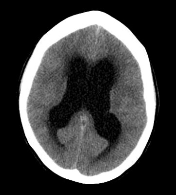
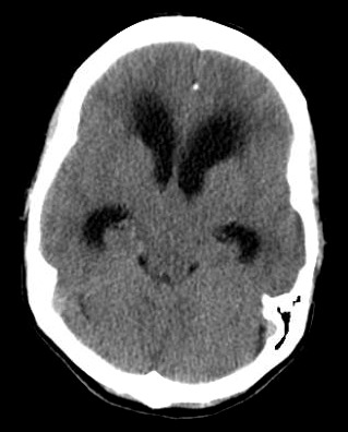
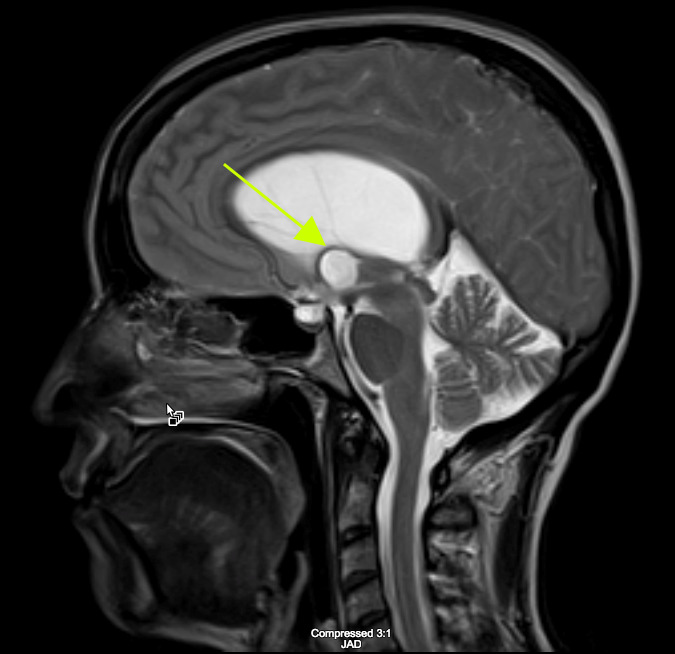
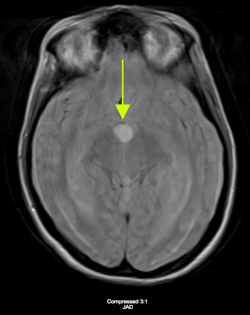
A Caucasian female in her late forties presented to the Emergency Department (ED) with headache, ataxia, and mental status changes. A CT brain demonstrated dilated lateral ventricles with transependymal edema. An MRI of the brain demonstrated marked obstructive hydrocephalus from an obstructing colloid cyst at the level of her Foramen of Monro. The patient was transferred to a tertiary care center for neurosurgical removal of the cyst. Three months later, the patient was doing well and had resumed all activities of daily living without any residual neurological deficits. The goal of this case report is to educate readers on this atypical presentation of hydrocephalus, its symptomatology, and management to allow physicians to be more comfortable in making treatment decisions.
Keywords: colloid cyst; emergency neurology; foramen of monro; hydrocephalus.
Figures
Similar articles
-
Colloid Cyst Presenting With Severe Headache and Bilateral Leg Weakness: Case Report and Review.Cureus. 2023 Nov 24;15(11):e49347. doi: 10.7759/cureus.49347. eCollection 2023 Nov. Cureus. 2023. PMID: 38143674 Free PMC article.
-
Third Ventricle Cavernous Malformation and Obstructive Hydrocephalus Thought to Be a Colloid Cyst.World Neurosurg. 2021 Jan;145:315-319. doi: 10.1016/j.wneu.2020.09.136. Epub 2020 Oct 1. World Neurosurg. 2021. PMID: 33010503
-
Third ventricular colloid cyst causing acute hydrocephalus during early pregnancy: Clinical lessons from a case.Surg Neurol Int. 2021 Feb 10;12:54. doi: 10.25259/SNI_724_2020. eCollection 2021. Surg Neurol Int. 2021. PMID: 33654557 Free PMC article.
-
[A case of colloid cyst of the third ventricle].No Shinkei Geka. 1988 Dec;16(13):1483-8. No Shinkei Geka. 1988. PMID: 3067109 Review. Japanese.
-
Giant, calcified colloid cyst of the lateral ventricle.J Clin Neurosci. 2016 Feb;24:6-9. doi: 10.1016/j.jocn.2015.05.044. Epub 2015 Sep 8. J Clin Neurosci. 2016. PMID: 26358201 Review.
References
-
- Spontaneous Regression of a Third Ventricle Colloid Cyst. Peters Sophie M., Daou Badih, Jabbour Pascal, Ladoux Alexandre, Georges Abi Lahoud. 2016World Neurosurg. 90(704):e19–22. - PubMed
-
- Macdonald R.L. In: Textbook of Neuro-oncology. Berger M.S., Prados M.D., editors. Elsevier Saunders; Philadelphia: Colloid Cysts in Children; p. 735.
-
- Bigner D.D., McLendon R.E., Bruner J.M. Russell and Rubinstein's Pathology of Tumors of the Nervous System. Hodder Headline Group; London:
-
- Natural history of colloid cysts of the third ventricle. Beaumont T.L., Limbrick Jr. D.D. , Rich K.M., Wippold F.J. 2nd, Dacey Jr. R.G. 2016J Neurosurg. 125(6):1–11. - PubMed
-
- Familial fatal and near-fatal third ventricle colloid cysts. Stoodley M.A., Nguyen T.P., Robbins P. 1999Aust N Z J Surg. 69(10):733–6. - PubMed
Publication types
LinkOut - more resources
Full Text Sources