

Effects of Ageing on Aortic Circulation During Atrial Fibrillation; a Numerical Study on Different Aortic Morphologies
- PMID: 33655419
- PMCID: PMC8455405
- DOI: 10.1007/s10439-021-02744-9
Effects of Ageing on Aortic Circulation During Atrial Fibrillation; a Numerical Study on Different Aortic Morphologies
Abstract
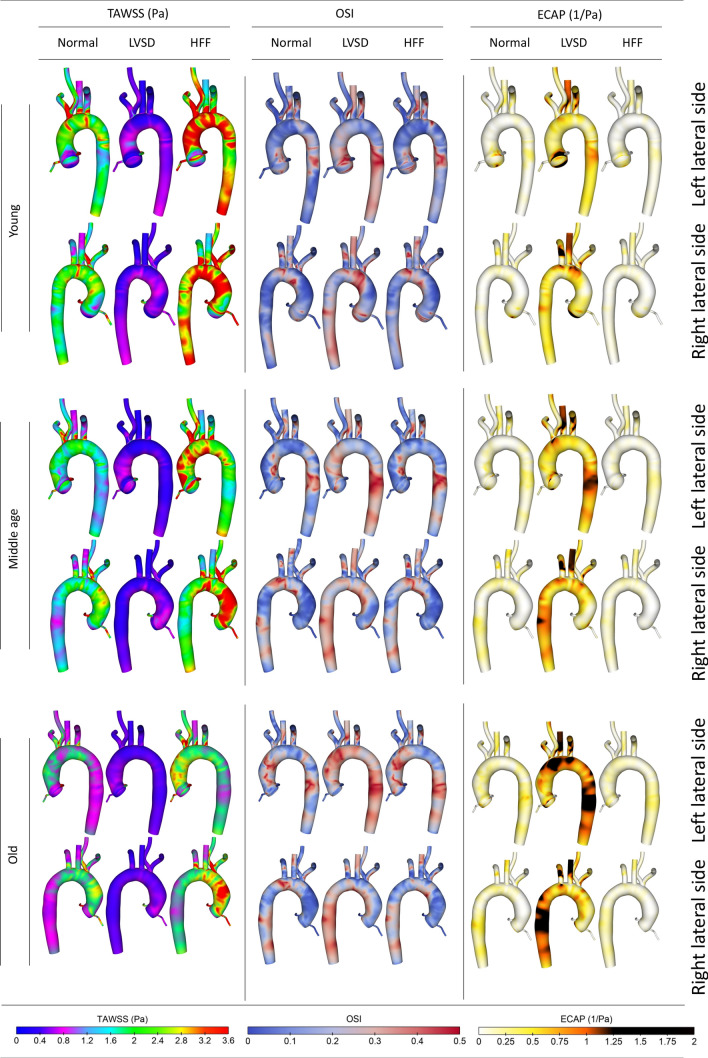
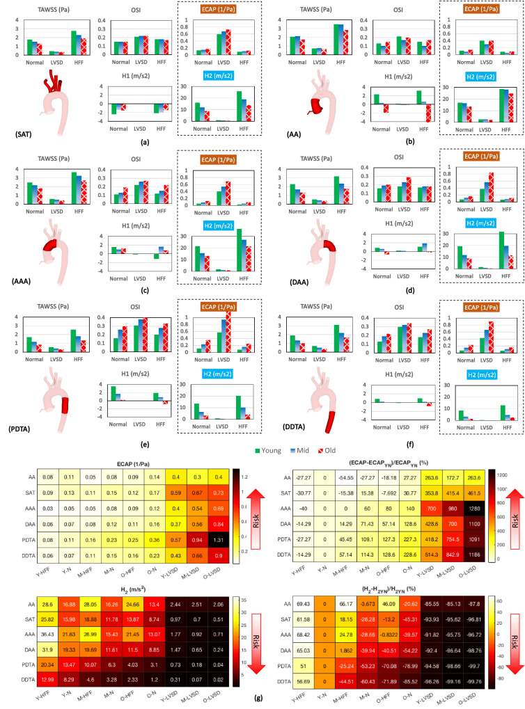
Atrial fibrillation (AF) can alter intra-cardiac flow and cardiac output that subsequently affects aortic flow circulation. These changes may become more significant where they occur concomitantly with ageing. Aortic ageing is accompanied with morphological changes such as dilation, lengthening, and arch unfolding. While the recognition of AF mechanism has been the subject of numerous studies, less focus has been devoted to the aortic circulation during the AF and there is a lack of such investigation at different ages. The current work aims to address the present gap. First, we analyse aortic flow distribution in three configurations, which attribute to young, middle and old people, using geometries constructed via clinical data. We then introduce two transient inlet flow conditions representative of key AF-associated defects. Results demonstrate that both AF and ageing negatively affect flow circulation. The main consequence of concomitant occurrence is enhancement of endothelial cell activation potential (ECAP) throughout the vascular domain, mainly at aortic arch and descending thoracic aorta, which is consistent with some clinical observations. The outcome of the current study suggests that AF exacerbates the vascular defects occurred due to the ageing, which increases the possibility of cardiovascular diseases per se.
Keywords: Ageing; Aorta; Atrial fibrillation; Cardiovascular diseases; Computational fluid dynamics; Haemodynamic metrics.
© 2021. The Author(s).
Figures
Similar articles
-
Numerical Study of Atrial Fibrillation Effects on Flow Distribution in Aortic Circulation.Ann Biomed Eng. 2020 Apr;48(4):1291-1308. doi: 10.1007/s10439-020-02448-6. Epub 2020 Jan 14. Ann Biomed Eng. 2020. PMID: 31938982 Free PMC article.
-
Role of the vessel morphology on the lenticulostriate arteries hemodynamics during atrial fibrillation: A CFD-based multivariate regression analysis.Comput Methods Programs Biomed. 2024 Sep;254:108303. doi: 10.1016/j.cmpb.2024.108303. Epub 2024 Jun 24. Comput Methods Programs Biomed. 2024. PMID: 38943985
-
Numerical analysis of hemodynamic changes in the left atrium due to atrial fibrillation.J Biomech. 2015 Feb 5;48(3):472-8. doi: 10.1016/j.jbiomech.2014.12.025. Epub 2014 Dec 16. J Biomech. 2015. PMID: 25547024
-
Imaging Insights on the Aorta in Aging.Circ Cardiovasc Imaging. 2018 Apr;11(4):e005617. doi: 10.1161/CIRCIMAGING.117.005617. Circ Cardiovasc Imaging. 2018. PMID: 29653929 Free PMC article. Review.
-
Advance in the application of 4-dimensional flow MRI in atrial fibrillation.Magn Reson Imaging. 2025 Jan;115:110254. doi: 10.1016/j.mri.2024.110254. Epub 2024 Oct 12. Magn Reson Imaging. 2025. PMID: 39401601 Review.
Cited by
-
Cerebral hemodynamics during atrial fibrillation: Computational fluid dynamics analysis of lenticulostriate arteries using 7 T high-resolution magnetic resonance imaging.Phys Fluids (1994). 2022 Dec;34(12):121909. doi: 10.1063/5.0129899. Epub 2022 Dec 29. Phys Fluids (1994). 2022. PMID: 36776539 Free PMC article.
-
Modulating activity of PVN neurons prevents atrial fibrillation induced circulation dysfunction by electroacupuncture at BL15.Chin Med. 2023 Oct 17;18(1):135. doi: 10.1186/s13020-023-00841-6. Chin Med. 2023. PMID: 37848944 Free PMC article.
-
Patient-Specific Haemodynamic Analysis of Virtual Grafting Strategies in Type-B Aortic Dissection: Impact of Compliance Mismatch.Cardiovasc Eng Technol. 2024 Jun;15(3):290-304. doi: 10.1007/s13239-024-00713-6. Epub 2024 Mar 4. Cardiovasc Eng Technol. 2024. PMID: 38438692 Free PMC article.
-
The Differences of Quantitative Flow Ratio in Coronary Artery Stenosis with or without Atrial Fibrillation.J Interv Cardiol. 2023 Oct 13;2023:7278343. doi: 10.1155/2023/7278343. eCollection 2023. J Interv Cardiol. 2023. PMID: 37868769 Free PMC article.
-
Multi-Objective Optimisation of a Novel Bypass Graft with a Spiral Ridge.Bioengineering (Basel). 2023 Apr 19;10(4):489. doi: 10.3390/bioengineering10040489. Bioengineering (Basel). 2023. PMID: 37106676 Free PMC article.
References
-
- Barth TJ, Jespersen DC. The design and application of upwind schemes on unstructured meshes. 1989 doi: 10.2514/6.1989-366. - DOI
-
- Benim AC, Nahavandi A, Assmann A, Schubert D, Feindt P, Suh SH. Simulation of blood flow in human aorta with emphasis on outlet boundary conditions. Appl. Math. Model. 2011;35:3175–3188. doi: 10.1016/j.apm.2010.12.022. - DOI
Publication types
MeSH terms
Grants and funding
LinkOut - more resources
Full Text Sources
Other Literature Sources
Medical
Miscellaneous
