. 2021 Mar 3;10(1):8.
doi: 10.1186/s13619-020-00071-3.

RhoA/Rock activation represents a new mechanism for inactivating Wnt/β-catenin signaling in the aging-associated bone loss

Affiliations

RhoA/Rock activation represents a new mechanism for inactivating Wnt/β-catenin signaling in the aging-associated bone loss

Wei Shi et al. Cell Regen. .

Erratum in

Abstract

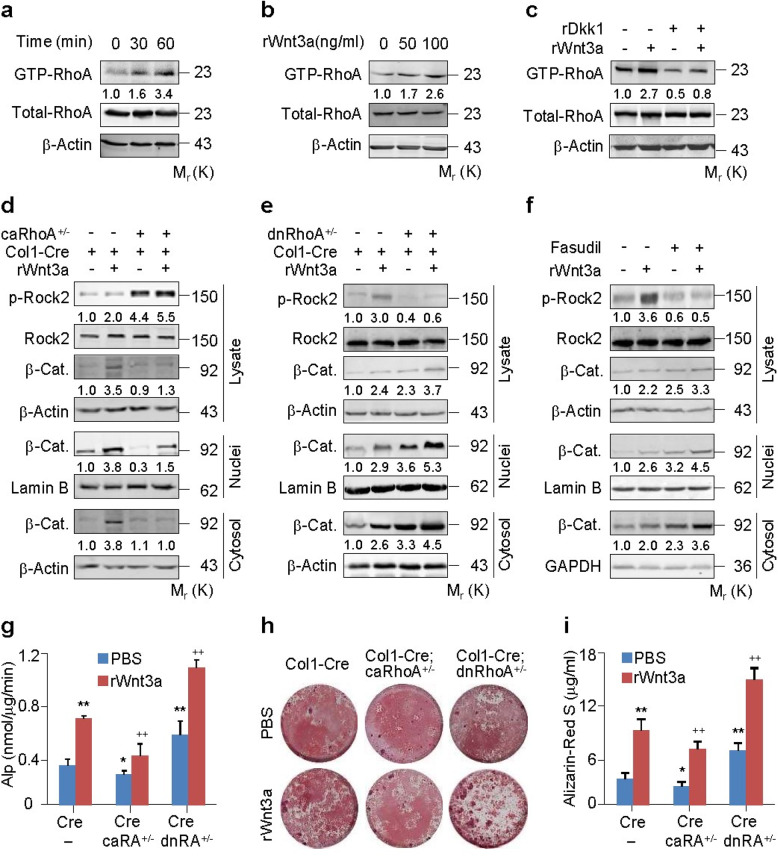
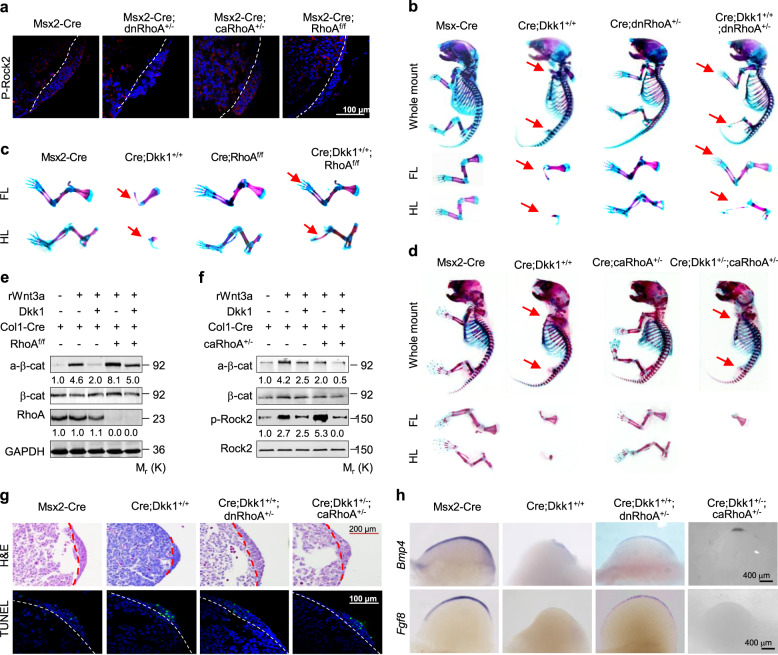
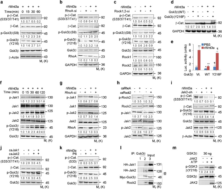
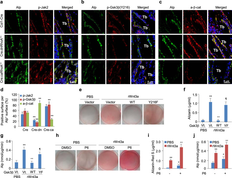
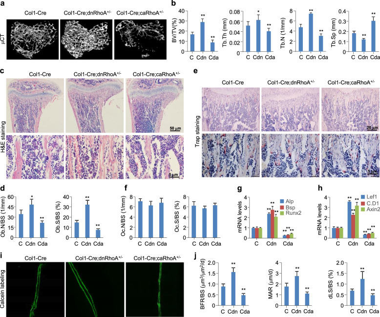
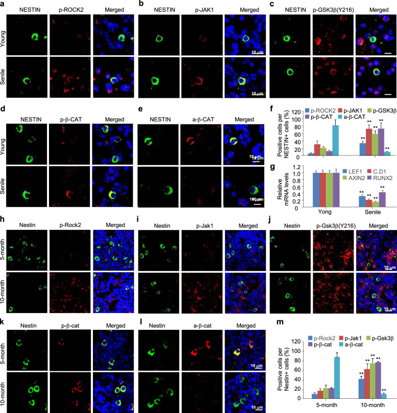
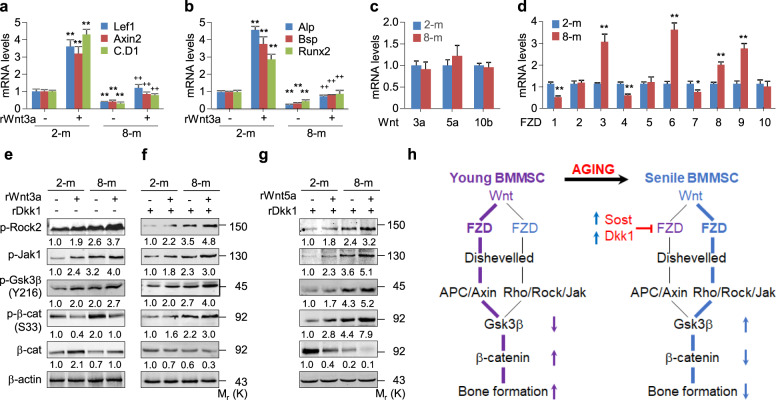
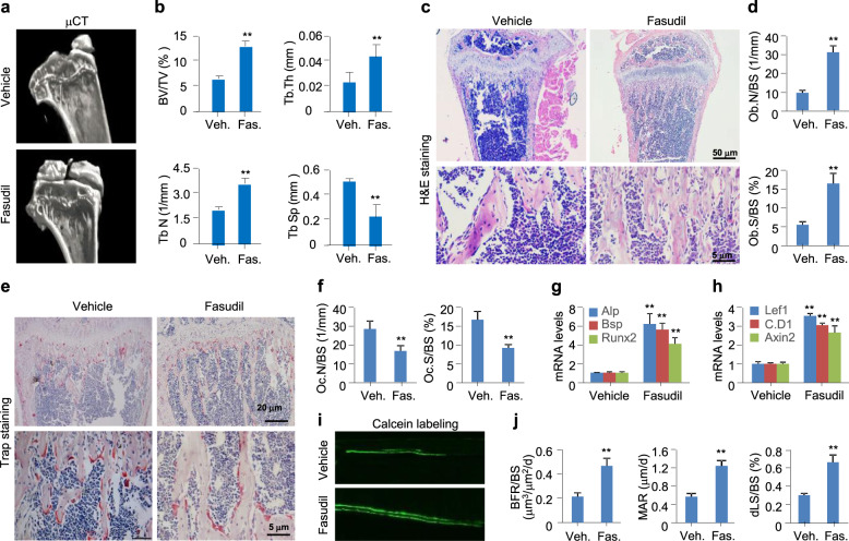
The Wnt/β-catenin signaling pathway appears to be particularly important for bone homeostasis, whereas nuclear accumulation of β-catenin requires the activation of Rac1, a member of the Rho small GTPase family. The aim of the present study was to investigate the role of RhoA/Rho kinase (Rock)-mediated Wnt/β-catenin signaling in the regulation of aging-associated bone loss. We find that Lrp5/6-dependent and Lrp5/6-independent RhoA/Rock activation by Wnt3a activates Jak1/2 to directly phosphorylate Gsk3β at Tyr216, resulting in Gsk3β activation and subsequent β-catenin destabilization. In line with these molecular events, RhoA loss- or gain-of-function in mouse embryonic limb bud ectoderms interacts genetically with Dkk1 gain-of-function to rescue the severe limb truncation phenotypes or to phenocopy the deletion of β-catenin, respectively. Likewise, RhoA loss-of-function in pre-osteoblasts robustly increases bone formation while gain-of-function decreases it. Importantly, high RhoA/Rock activity closely correlates with Jak and Gsk3β activities but inversely correlates with β-catenin signaling activity in bone marrow mesenchymal stromal cells from elderly male humans and mice, whereas systemic inhibition of Rock therefore activates the β-catenin signaling to antagonize aging-associated bone loss. Taken together, these results identify RhoA/Rock-dependent Gsk3β activation and subsequent β-catenin destabilization as a hitherto uncharacterized mechanism controlling limb outgrowth and bone homeostasis.

Keywords: Bone; Limb bud; RhoA; Rock; Wnt; β-Catenin.


Conflict of interest statement

The authors declare no competing interests.

Figures

Fig. 1
Fig. 1
RhoA/Rock constraints Wnt/β-catenin signaling and osteoblastic differentiation. a-c RhoA activation assays in primary murine calvarial osteoblasts (PMCOBs) stimulated with rWnt3a at 100 ng/ml or the indicated concentrations for the indicated times or 60 min in the presence or absence of recombinant Dkk1 (rDkk1) at 100 ng/ml. d, e Western analyses of β-catenin in cytosolic and nuclear fractions of PMCOBs with the indicated genotypes of Col1-Cre (Cre), Col1-Cre;caRhoA+/− (Cre;caRhoA+/−) or Col1-Cre;dnRhoA+/− (Cre;dnRhoA+/−), and in the presence or absence of rWnt3a for 3 h. f Western analyses of β-catenin (β-cat) in cytosolic and nuclear fractions of PMCOBs treated with or without Fasudil at 20 μM and stimulated with or without rWnt3a for 3 h. g-i Alp activity and mineralization nodule formation assays and their quantification in PMCOBs with the indicated genotypes and stimulated with or without rWnt3a at 100 ng/ml for 48 h and 21 d, respectively. Mean ± SEM, *p < 0.05, **,++p < 0.01, n = 4, Tukey-Kramer multiple comparisons test
Fig. 2
Fig. 2
RhoA interacts genetically with Dkk1 in the limb bud ectoderm of mouse embryos. a Representative immunostaining for p-Rock2 in the E10.5 limb buds with the indicated genotypes. White dot lines separate the apical ectodermal ridge (AER) from the zone of polarizing activity (ZPA). b-d Skeletons and/or limbs of E16.5 embryos with the indicated genotypes. e, f Western analyses in PMCOBs with the indicated genotypes and stimulated with or without rWnt3a at 100 ng/ml for 1 h in the presence or absence of rDkk1 at 100 ng/ml. Phosphorylated proteins were normalized to their total amounts, respectively. g, h H&E and TUNEL staining in E10.5 forelimb bud sections (g) and whole-mount in situ hybridization of E10.5 forelimb buds (h). Dot lines separate the AER from the ZPA. Ventral view for all limb buds, anterior to the lower and posterior to the upper. FL: forelimb, HL: hindlimb
Fig. 3
Fig. 3
RhoA/Rock activates Jak1/2 and Gsk3β to destabilize β-catenin. a-c Western analyses in C3H10T1/2 cells transfected with or without RhoA-si or Rock1 + Rock2 siRNA (Rock1,2-si) and treated with or without rWnt3a at 100 ng/ml for the indicated time or 1 h. d, e Western or Lef1-luciferase expression analyses in C3H10T1/2 cells transfected with Gsk3β variants and treated with rWnt3a for 6 or 48 h, respectively. f-k Western analyses in C3H10T1/2 cells transfected with RhoA-si, caRhoA, caRock2, caJak1/2, infected with lentiviral Jak2-shRNA (Jak2-sh), or treated with P6 at 50 nM, followed by incubation with rWnt3a for the indicated times or 1 h. l Co-immunoprecipitation by using IgG1 or Gsk3β antibody in 293 cells transfected with HA-Jak1/2 and Myc-Gsk3β. m In vitro phosphorylation of GSK3β protein by active JAK2 in kinase assay buffer with or without ATP. Phosphorylated proteins were normalized to their total amounts, respectively. Mean ± SD, *, ¶ p < 0.05, **, ++p < 0.01, n = 4, Tukey-Kramer multiple comparisons test
Fig. 4
Fig. 4
RhoA/Rock activates Jak and GSK3β to destabilize β-catenin in osteoblastic differentiation in response to Wnt3a. a-c Images co-stained Alp with p-Jak2, p-Tyr216-Gsk3β or active β-catenin in proximal tibia (Tb) sections of 2-month-old Col1-Cre, Col1-Cre;dnRhoA+/−, or Col1-Cre;caRhoA+/− mice. d The percentages of p-Jak2+, p-Tyr216-Gsk3β+, or active β-catenin+ surface in Alp+ surface were quantified. Mean ± SEM, **p < 0.01, n = 6, Tukey-Kramer multiple comparisons test. e-j Alizarin Red S staining and quantification or Alp activity assays in C3H10T1/2 cells infected with vector-, Gsk3β(WT)- or Gsk3β(Y216F)-expressing lentiviruses or treated with vehicle or P6 at 50 nM, and further cultured with or without 100 ng/ml of rWnt3a for 21 days or 48 h, respectively. Mean ± SD, *, p < 0.05, **,++p < 0.01, n = 3 ~ 5, Tukey-Kramer multiple comparisons test
Fig. 5
Fig. 5
RhoA loss- or gain-of-function affects the bone mass. Tibias from 8-week-old male mice with the genotypes of Col1-Cre (C), Col1-Cre;dnRhoA+/− (Cdn) or Col1-Cre;caRhoA+/− (Cda) were harvested for the following analyses. a, b Representative μCT images and parameters including BV/TV, Tb.Th, Tb.N, and Tb.Sp for the proximal tibias. c, d Representative images of H&E staining and quantification of Ob.N/BS and Ob.S/BS for the proximal tibia sections. e, f Representative images of Trap staining and quantification of Oc.N/BS and Oc.S/BS for the proximal tibia sections. g, h qPCR analyses for Alp, Bsp, Runx2, Lef1, Cyclin D1 (C.D1), and Axin2 mRNA levels in femurs. i, j Dynamic bone histomorphometry and quantification for BFR/BS, MAR, and dLS/BS. Mean ± SEM, *p < 0.05, **p < 0.01, n = 8, Tukey-Kramer multiple comparisons test
Fig. 6
Fig. 6
High RhoA/Rock/Jak1/Gsk3β activity is inversely correlated with Wnt/β-catenin signaling activity in the BMMSCs from elderly subjects. a-e, h-l Immunofluorescence images co-stained Nestin with p-Rock2, p-Tyr1022-Jak1, p-Tyr216-Gsk3β, p-S33-β-catenin (p-β-cat), or non-p-Ser45-β-catenin (a-β-cat) in bone marrow smears from either elderly men (n = 4) with femoral neck fractures and type II osteoporosis or young males (n = 3) with traumatic fractures and normal bone volumes, and in the proximal tibia sections of 5- or 10-month-old mice. f, m The percentages of p-Rock2+, p-Tyr1022-Jak1+, p-Tyr216-Gsk3β+, p-β-cat+, or a-β-cat+ cells in Nestin+ cells. g Quantitative RT-PCR analyses for LEF1, Cyclin D1 (C.D1), AXIN2, and RUNX2 mRNA levels in the BMMSCs isolated form the elderly or young men. Mean ± SEM, **p < 0.01, Student’s t test
Fig. 7
Fig. 7
FZD receptors and Dkk1 coordinate RhoA/Rock activation to destabilize β-catenin in the BMMSCs from elderly mice. a-d Quantitative RT-PCR analyses for the indicated mRNA levels of BMMSCs isolated form 2- or 8-month-old mice and treated with or without rWnt3a at 100 ng/ml for 48 h. Mean ± SD, **p < 0.01, n = 6, Tukey-Kramer multiple comparisons test. e-g Western blotting analyses in the BMMSCs isolated form 2- or 8-month-old mice and treated with or without rWnt3a/rWnt5a at 100 ng/ml in the presence or absence of rDkk1 at 100 ng/ml for 3 h. h An integrated working model of β-catenin signaling mediated by RhoA/Rock in the regulation of aging-associated bone loss. In the BMMSCs from young subjects, APC/Axin/Gsk3β signaling mediated by FZD1, FZD4, and FZD7 overwhelms the RhoA/Rock/Jak/Gsk3β signaling mediated by FZD3, FZD6, and FZD8 to stabilize β-catenin and, in turn, enhance bone formation. However, in the BMMSCs from elderly subjects, the activation of RhoA/Rock/Jak/Gsk3β signaling mediated by FZD3, FZD6, and FZD8, in combination with the inactivation of APC/Axin/Gsk3β signaling mediated by Dkk1, Sost, and FZD1, FZD4, and FZD7 results in the destabilization of β-catenin and the subsequent attenuation of bone formation
Fig. 8
Fig. 8
Pharmacological inhibition of Rock by Fasudil antagonizes aging-associated bone loss. 8-month-old male mice were orally administrated with vehicle (Veh.) or Fasudil (Fas.) at 100 mg/kg once daily for 2 months. Tibias were harvested for the following analyses. a, b Representative μCT images and parameters including BV/TV, Tb.Th, Tb.N, and Tb.Sp for the proximal tibias. c, d Representative images of H&E staining and quantification of Ob.N/BS and Ob.S/BS for the proximal tibia sections. e, f Representative images of Trap staining and quantification of Oc.N/BS and Oc.S/BS for the proximal tibia sections. g, h qPCR analyses for Alp, Bsp, Runx2, Lef1, Cyclin D1 (C.D1), and Axin2 mRNA levels in femurs. (i, j) Dynamic bone histomorphometry and quantification for BFR/BS, MAR, and dLS/BS. Mean ± SEM, *p < 0.05, **p < 0.01, n = 8, Tukey-Kramer multiple comparisons test

References

    1. Adam S, Simon N, Steffen U, Andes FT, Scholtysek C, Müller DIH, et al. JAK inhibition increases bone mass in steady-state conditions and ameliorates pathological bone loss by stimulating osteoblast function. Sci Transl Med. 2020;12:eaay4447. doi: 10.1126/scitranslmed.aay4447. - DOI - PubMed
    1. AlMuraikhi N, Ali D, Alshanwani A, Vishnubalaji R, Manikandan M, Atteya M, et al. Stem cell library screen identified ruxolitinib as regulator of osteoblastic differentiation of human skeletal stem cells. Stem Cell Res Ther. 2018;9:319. doi: 10.1186/s13287-018-1068-x. - DOI - PMC - PubMed
    1. Bafico A, Liu G, Yaniv A, Gazit A, Aaronson SA. Novel mechanism of Wnt signalling inhibition mediated by Dickkopf-1 interaction with LRP6/arrow. Nat Cell Biol. 2001;3:683–686. doi: 10.1038/35083081. - DOI - PubMed
    1. Barrow JR, Thomas KR, Boussadia-Zahui O, Moore R, Kemler R, Capecchi MR, et al. Ectodermal Wnt3/beta-catenin signaling is required for the establishment and maintenance of the apical ectodermal ridge. Genes Dev. 2003;17:394–409. doi: 10.1101/gad.1044903. - DOI - PMC - PubMed
    1. Bishop AL, Hall A. Rho GTPases and their effector proteins. Biochem J. 2000;348:241–255. doi: 10.1042/bj3480241. - DOI - PMC - PubMed

LinkOut - more resources