. 2021 May 3;218(5):e20201452.
doi: 10.1084/jem.20201452.

Vincristine-induced peripheral neuropathy is driven by canonical NLRP3 activation and IL-1β release

Affiliations

Vincristine-induced peripheral neuropathy is driven by canonical NLRP3 activation and IL-1β release

Hana Starobova et al. J Exp Med. .

Abstract

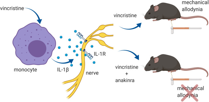
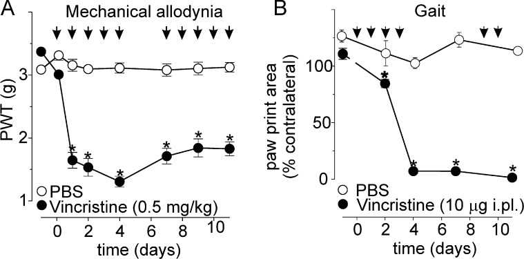
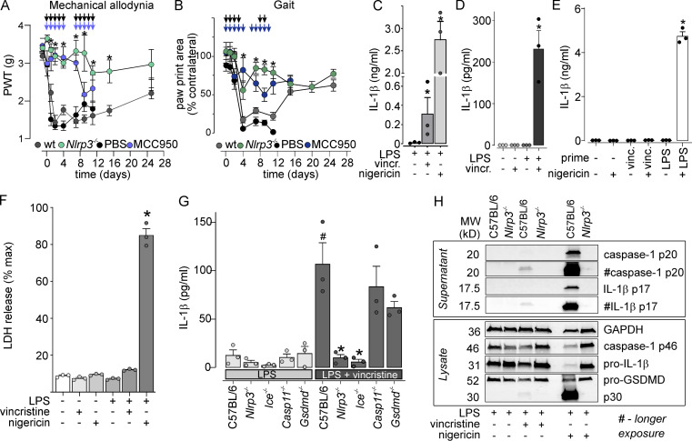
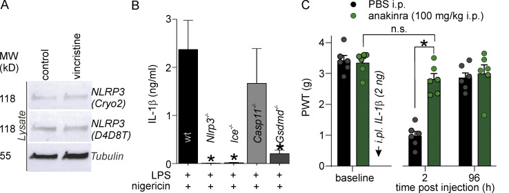
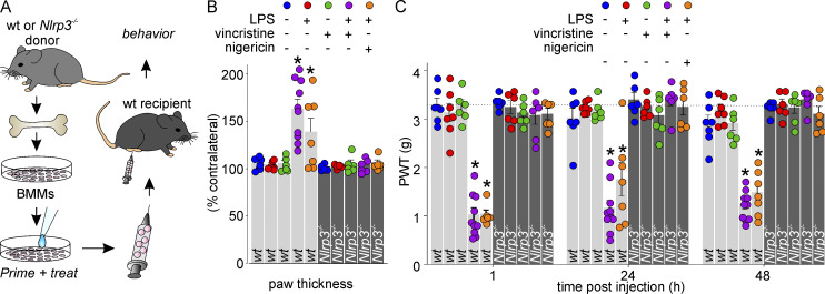
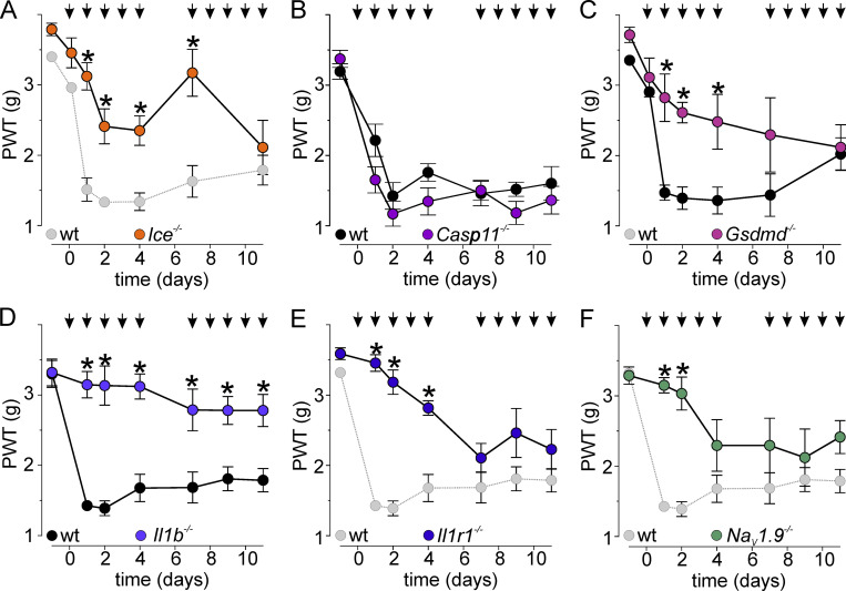
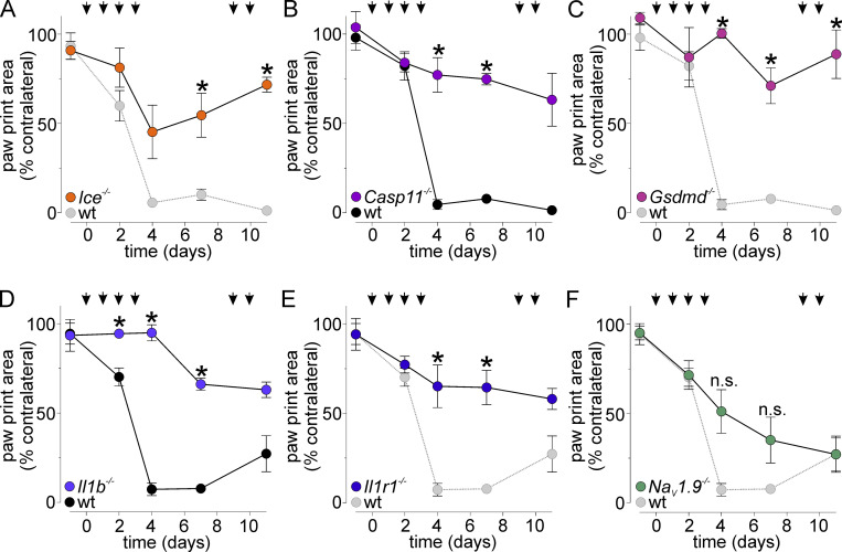
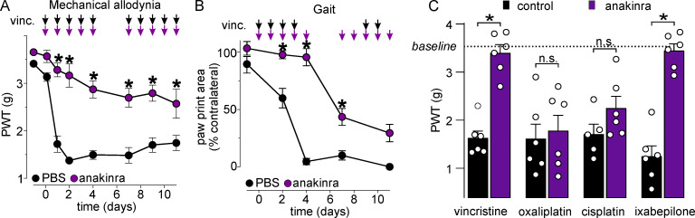
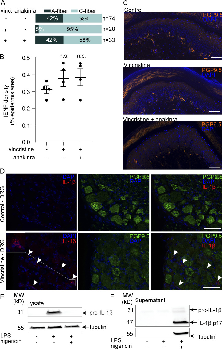
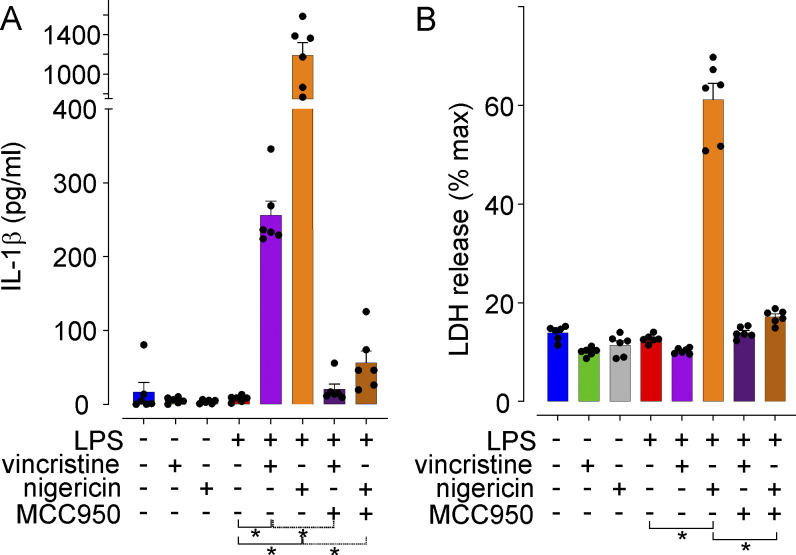
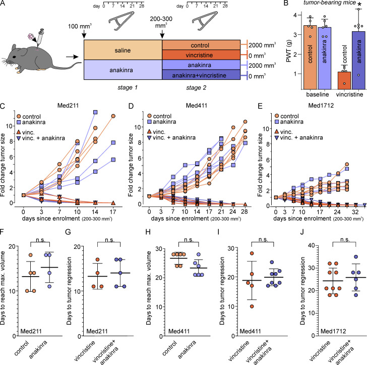
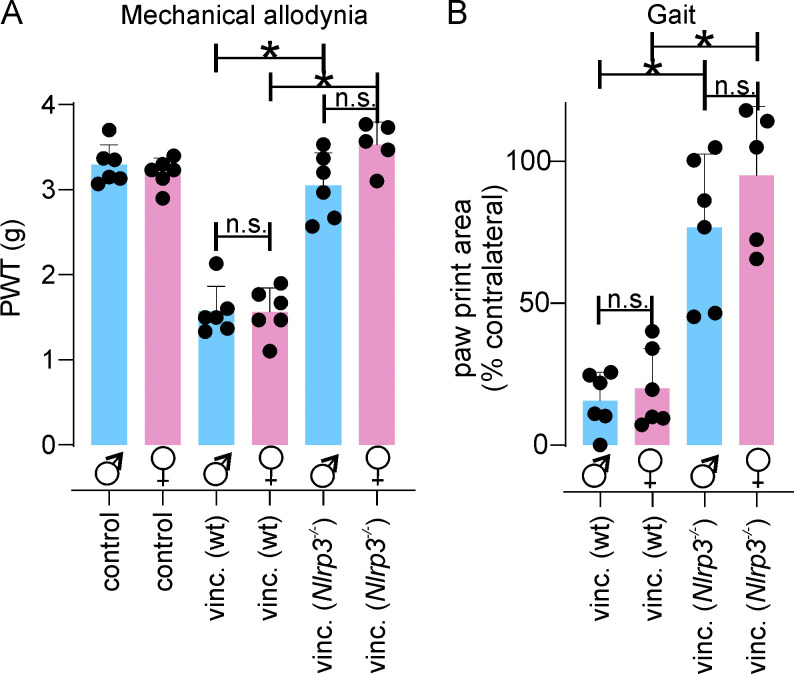
Vincristine is an important component of many regimens used for pediatric and adult malignancies, but it causes a dose-limiting sensorimotor neuropathy for which there is no effective treatment. This study aimed to delineate the neuro-inflammatory mechanisms contributing to the development of mechanical allodynia and gait disturbances in a murine model of vincristine-induced neuropathy, as well as to identify novel treatment approaches. Here, we show that vincristine-induced peripheral neuropathy is driven by activation of the NLRP3 inflammasome and subsequent release of interleukin-1β from macrophages, with mechanical allodynia and gait disturbances significantly reduced in knockout mice lacking NLRP3 signaling pathway components, or after treatment with the NLRP3 inhibitor MCC950. Moreover, treatment with the IL-1 receptor antagonist anakinra prevented the development of vincristine-induced neuropathy without adversely affecting chemotherapy efficacy or tumor progression in patient-derived medulloblastoma xenograph models. These results detail the neuro-inflammatory mechanisms leading to vincristine-induced peripheral neuropathy and suggest that repurposing anakinra may be an effective co-treatment strategy to prevent vincristine-induced peripheral neuropathy.


Conflict of interest statement

Disclosures: K. Schroder reported "other" from Inflazome Ltd outside the submitted work; in addition, K. Schroder had a patent to PCT/EP2017/053498 licensed (Inflazome Ltd), a patent to PCT/IB2017/053059 licensed (Inflazome Ltd), and a patent to PCT/AU2016/050103 licensed (Inflazome Ltd); served on the Scientific Advisory Board of Inflazome in 2016-2017; and serves as a consultant to Quench Bio, USA and Novartis, Switzerland. No other disclosures were reported.

Figures

None
Graphical abstract
Figure 1.
Figure 1.
A murine model of vincristine-induced neuropathy recapitulates sensory and gait abnormalities. (A) Vincristine (black circles; n = 6) causes a decrease of mechanical PWTs in C57BL/6J mice compared with PBS-treated animals (white circles; n = 6). (B) Vincristine (black circles; n = 6) causes gait abnormalities, evidenced by a decrease of paw print area (cm2) relative to the contralateral control (PBS, white circles). Black arrows in A and B indicate the time points of vincristine administration. Statistical significance was determined by repeated measures two-way ANOVA with Sidak’s multiple comparisons test; data are shown as mean ± SEM. *, P < 0.05.
Figure S1.
Figure S1.
Effect of vincristine on weight gain, motor performance, and heat sensitivity in C57BL/6 mice. (A–C) Vincristine (0.5 mg/kg i.p.) does not affect weight gain (P > 0.05 by two-way repeated measures ANOVA) in either males or females (A), and does not affect motor performance (B and C), assessed using the parallel rod floor apparatus. Both distance traveled (m; B) and foot slips/m (C) were unchanged (P > 0.05 by unpaired t test) in vincristine-treated animals compared with control. (D) Vincristine (0.5 mg/kg i.p.) does not cause heat allodynia, with PWTs unchanged compared with PBS-treated animals (P > 0.05 by two-way repeated measures ANOVA). All data are shown as mean ± SEM; n ≥5 for all groups as indicated.
Figure 2.
Figure 2.
VIPN is driven by infiltrating macrophages. (A–I) Vincristine (vinc) causes infiltration of F4/80+ cells (arrows; brown due to diaminobenzidine chromogen) into the sciatic nerve (A–C) and DRGs (D–F), but not spinal cord (G–I). Representative IHC images of sciatic nerve (A and B), DRG (D and E), and spinal cord (G and H) sections from C57BL/6J mice. Scale bar: 50 µm. (J–L) Depletion of macrophages by liposomal clodronate was confirmed by quantitative anti-F4/80+ staining of spleen (J and K, representative IHC images; scale bar: 200 µm). The inset in J and K is a magnified view showing F4/80+ cells. (M) Compared with PBS-treated animals, treatment with liposomal clodronate (lip. clodronate; 50 µg/g i.p.; 24 h) significantly decreased the number of F4/80+ cells infiltrating the sciatic nerve of animals after vincristine administration. (N) Depletion of granulocytes in circulating blood after treatment with anti-Ly6G antibody (200 µg i.p.) compared with the isotype control antibody (200 µg); confirmed by flow cytometry. (O) Depletion of macrophages with liposomal clodronate (black bars), but not granulocytes (light gray bars), reverses development of vincristine-induced mechanical allodynia compared with vehicle (white bars) and isotype antibody control (dark gray bars). Statistical significance was determined by unpaired two-tailed t test (C, F, I, L, M, and N) and two-way ANOVA with Tukey’s multiple comparisons test (O). Data are shown as mean ± SEM; n = 3–7 for all groups as indicated. *, P < 0.05; **, P < 0.01.
Figure 3.
Figure 3.
Vincristine leads to activation of the NLRP3-inflammasome and IL-1β release. (A) Mechanical allodynia induced by vincristine (vinc or vincr; 0.5 mg/kg i.p.) does not develop in Nlrp3−/− animals (green symbols) and is attenuated by the NLRP3 antagonist MCC950 (blue symbols; 20 mg/kg i.p.) compared with wt (dark gray symbols) and PBS-treated (black symbols) controls, respectively. Black arrows indicate vincristine administration schedule and colored arrows indicate MCC950 treatment schedule. n = 6 for all groups. (B) The decrease in paw print area induced by vincristine (10 µg i.pl.) is abolished in Nlrp3−/− animals (dark green) and after treatment with MCC950 (dark blue; 20 mg/kg i.p.) compared with wt (dark gray symbols) and PBS-treated (black symbols) controls, respectively. Black arrows indicate vincristine administration schedule and colored arrows indicate MCC950 treatment schedule. Statistical significance was determined by using two-way ANOVA with Tukey’s multiple comparisons test. All data are shown as mean ± SEM; n = 6 for all groups. *, P < 0.05. (C and D) Vincristine induces significant release of IL-1β in human (C) and mouse (D) LPS-primed macrophages. (E) Incubation of BMMs with vincristine does not prime the NLRP3 inflammasome. Nigericin (5 µM) treatment leads to release of IL-1β in LPS-primed, but not vincristine-primed, macrophages. (F) Vincristine treatment does not induce marked cell death, expressed as percentage of LDH release of 100% lysis control. (G) Vincristine-induced release of IL-1β is abolished in Nlrp3−/− and Ice−/−, but not in Casp11−/− or Gsdmd−/− LPS-primed BMMs. *, P < 0.05 cf. C57BL/6 (LPS);  #, P < 0.05 cf. C57BL/6 (LPS + vincristine). (H) Cleaved IL-1β (p17) and caspase-1 (p20) are found in supernatants of vincristine-treated C57BL6/J, but not Nlrp3−/−, LPS-primed BMMs. (C–G) Statistical significance was determined using one-way ANOVA. All data are shown as mean ± SEM; n = 3 for all groups. *, P < 0.05. MW, molecular weight.
Figure S2.
Figure S2.
Effect of vincristine and nigericin on the NLRP3 inflammasome in BMMs, and effects of anakinra on IL-1β-induced allodynia. (A) Vincristine treatment does not increase NLRP3 expression in macrophages, assessed by Western blotting using two anti-NLRP3 antibodies (Cryo2 and D4D8T). MW, molecular weight. (B) Nigericin-induced IL-1β release in LPS-primed C57BL6/J, Nlrp3−/−, Ice−/−, Casp11−/−, and Gsdmd−/− BMMs. Incubation time of Nigericin: 45 min. Statistical significance was determined using ordinary one-way ANOVA with multiple comparison. n = 3 independent experiments for all groups. (C) Decreased PWT induced by i.pl. injection of IL-1β (black bars) is prevented by treatment with anakinra (100 mg/kg i.p., green bars). n = 6 animals/group. Statistical significance was determined using ordinary two-way ANOVA with multiple comparison. All data are shown as mean ± SEM; *, P < 0.05. MW, molecular weight.
Figure 4.
Figure 4.
Intraplantar injection of vincristine-treated macrophages in naive animals causes mechanical allodynia. (A) Schematic illustrating the experimental design. BMMs isolated from WT or Nlrp3−/− animals were primed and treated in vitro, harvested, and administered to naive animals via i.pl. injection. (B) Intraplantar injection of LPS-primed macrophages treated with vincristine (purple) or nigericin (orange), but not untreated (blue), LPS-primed (red), unprimed vincristine-treated (green), or Nlrp3−/− macrophages (dark gray bars) causes unilateral paw swelling. n = 6–10 animals/group. (C) Intraplantar injection of LPS-primed macrophages treated with vincristine (purple) or nigericin (orange), but not untreated (blue), LPS-primed (red), or unprimed vincristine-treated (green) cells causes mechanical allodynia. Intraplantar injection of macrophages from Nlrp3−/− animals does not cause mechanical allodynia, irrespective of priming or treatment (dark gray bars). Dotted line indicates baseline PWT. n = 6–10 animals/group. Statistical significance was determined using one-way ANOVA. *, P < 0.05.
Figure 5.
Figure 5.
The canonical NLRP3 signaling pathway contributes to vincristine-induced mechanical allodynia. (A–E) Mechanical allodynia induced by treatment with vincristine (black arrows; 0.5 mg/kg i.p.) is attenuated in (A) Ice−/− animals (orange symbols; dotted line and light gray symbols are cohort controls also shown in Fig. 3 A); not attenuated in (B) Casp11−/− animals (purple symbols) relative to wt controls (black symbols); attenuated in (C) Gsdmd−/− animals (pink symbols; black symbols are cohort controls); and attenuated in (D) Il1b−/− animals relative to wt controls (black symbols) and (E) Il1r1−/− animals (dotted line and light gray symbols are cohort controls also shown in D). (F) In NaV1.9−/− animals, mechanical allodynia induced by vincristine (0.5 mg/kg; i.p.) is partially attenuated; dotted line and light gray symbols are cohort controls also shown in D. Statistical significance was determined using repeated measures two-way ANOVA with Sidak’s multiple comparisons test. All data are shown as mean ± SEM; n = 6 for all groups. *, P < 0.05.
Figure 6.
Figure 6.
NLRP3 signaling contributes to vincristine-induced gait disturbances. (A–E) Gait disturbances, evidenced by a decrease in the ipsilateral paw print area (shown as percentage of contralateral), induced by treatment with vincristine (black arrows; 10 µg i.pl.) are attenuated in (A) Ice−/− animals (orange symbols; dotted line and light gray symbols are cohort controls also shown in Fig. 3 B); (B) Casp11−/− animals (purple symbols) relative to wt controls (black symbols); (C) Gsdmd−/− animals (pink symbols; dotted line and light gray symbols are cohort controls also shown in B); (D) Il1b−/− animals relative to wt controls (black symbols); and (E) Il1r1−/− animals (dotted line and light gray symbols are cohort controls also shown in D). (F) In NaV1.9−/− animals, gait disturbances induced by vincristine are not significantly decreased; dotted line and light gray symbols are cohort controls also shown in D. Statistical significance was determined using repeated measures two-way ANOVA with Sidak’s multiple comparisons test. All data are shown as mean ± SEM; n = 6 for all groups. *, P < 0.05.
Figure 7.
Figure 7.
Anakinra treatment prevents the development of VIPN. (A) In animals receiving vincristine (vinc; 0.5 mg/kg i.p.), coadministration of anakinra (purple symbols; 100 mg/kg i.p.) fully prevents development of mechanical allodynia compared with animals treated with vehicle (black symbols; PBS i.p.). n = 6 animals/group. (B) Gait disturbances induced by vincristine are significantly (P < 0.05) delayed in animals treated with anakinra (purple symbols; 100 mg/kg i.p.) compared with animals treated with vehicle (black symbols; PBS i.p.). Black arrows indicate vincristine administration, and purple arrows indicate anakinra/PBS administration schedule. n = 6 animals/group. (C) Anakinra (100 mg/kg i.p., purple bars) reverses vincristine- and ixabepilone-, but not oxaliplatin- or cisplatin-induced mechanical allodynia. n = 6 animals/group. Statistical significance was determined using one-way (C) and repeated measures two-way ANOVA with Sidak’s multiple comparisons test (A and B). All data are shown as mean ± SEM. *, P < 0.05.
Figure S3.
Figure S3.
Effects of vincristine on peripheral nerve excitability, intraepidermal nerve fiber density, and IL-1β immunofluorescence in dorsal root ganglia. (A) The proportion of excitable myelinated A-fibers is reduced from 31/74 fibers in control animals (recordings from n = 5 animals) to 1/20 fibers after treatment with vincristine (vinc; recordings from n = 3 animals), and rescued (to 14/33 fibers) by anakinra (100 mg/kg i.p.; recordings from n = 3 animals). Excitable A- and C-fibers were classified by conduction velocity and quantified from single-fiber recordings using the murine skin-saphenous nerve preparation. (B and C) Vincristine treatment does not reduce the density of intraepidermal nerve fibers (IENFs; P > 0.05 by one-way ANOVA). (B) Area of pan-neuronal PGP9.5-positive immunofluorescence (percent epidermal area) from four to six nonconsecutive 50-µm skin sections taken from animals (n = 4/group) treated with PBS (control), vincristine (0.5 mg/kg i.p.), and vincristine + anakinra (0.5 mg/kg i.p./100 mg/kg i.p.) for 72 h. (C) Representative apotome images of IENFs in hind paw skin samples from control, vincristine-, and vincristine + anakinra-treated mice. Scale bar: 50 µm. (D) Representative immunofluorescence images of DRG sections (10 µm) from vehicle- and vincristine-treated mice stained with the pan-neuronal marker PGP9.5 (green) and anti–IL-1β (red; AF-401NA; R&D Systems). Arrowheads: IL-1β–positive cells are observed in close proximity to PGP9.5-positive DRG neuron cell bodies. Scale bar: 50 µm. (E and F) Anti–IL-1β antibody AF-401NA detects uncleaved pro–IL-1β in cell lysates from LPS-primed macrophages (E), as well as cleaved, mature IL-1β secreted into cell supernatants by LPS-primed, nigericin-treated macrophages (F). MW, molecular weight.
Figure S4.
Figure S4.
Activation of the NLRP3 inflammasome in macrophages from NSG mice. (A) LPS-primed, but not unprimed, macrophages isolated from NSG mice respond to both vincristine and nigericin with an increase in secreted IL-1β that is reversed by treatment with the NLRP3 inhibitor MCC950. (B) Nigericin treatment induced pyroptosis, evidenced by an increase in LDH release, in LPS-primed macrophages from NSG mice. All data are shown as mean ± SEM; n = 6 replicates for all groups. Statistical significance was determined by one-way ANOVA. *, P < 0.05.
Figure 8.
Figure 8.
Anakinra treatment does not affect tumor progression or chemotherapy efficacy. (A) Detailed time schedule of drug administration in Med-211FH, Med-411FH, and Med-1712FH medulloblastoma PDX models. Mice were injected with Med-211FH, Med-411FH, and Med-1712FH medulloblastoma cells and tumor volume measurements were taken biweekly. Anakinra (light purple) or saline (light orange) treatment commenced when s.c. tumors reached 100 mm3 in size (stage 1). Vincristine (dark orange), anakinra (light purple), saline (light orange), or anakinra + vincristine (dark purple) injections commenced when s.c. tumors reached 300 mm3 in size (stage 2) and treatment was administered for 4 wk daily or until maximum tumor size was reached (2,000 mm3). (B) Anakinra (100 mg/kg i.p., purple bars) reverses vincristine-induced mechanical allodynia in tumor-bearing NSG mice. (C–J) Effect of anakinra on tumor growth and vincristine-induced complete tumor regression in Med-211FH, Med-411FH, and Med-1712FH medulloblastoma PDX models. (C–F and H) Daily treatment with anakinra (light purple) had no effect on time to reach maximum tumor size (2,000 mm3) or tumor growth in animals with an s.c. implanted Med-211FH or Med-411FH tumor compared with controls (light orange symbols). There was also no effect on tumor growth in animals bearing Med-1712FH tumors. (C–E) Effect of anakinra on vincristine-induced complete tumor regression in Med-211FH, Med-411FH, and Med-1712FH medulloblastoma PDX models. (C, D, E, G, I, and J) Daily treatment with anakinra (dark purple symbols) had no effect on the ability of vincristine to induce regression of Med-211FH (G), Med-411FH (I), or Med-1712FH (J) tumors compared with control animals treated only with vincristine (dark orange symbols). All data are shown as mean ± SEM; n ≥ 4 animals/group as indicated. *, P < 0.05.
Figure S5.
Figure S5.
Vincristine-induced neuropathy in male and female C57BL/6 and Nlrp3/ mice. (A and B) Vincristine (vinc)-induced mechanical allodynia (A) and gait disturbances (B) develop in both male (blue) and female (pink) animals. Both male and female Nlrp3−/− animals are protected from vincristine-induced mechanical allodynia (A) and gait disturbances (B). All data are shown as mean ± SEM; n ≥5 for all groups. Statistical significance was determined by one-way ANOVA with Sidak’s post-test. *, P < 0.05.

Comment in

Similar articles

Cited by

References

    1. Abbate, A., Salloum F.N., Vecile E., Das A., Hoke N.N., Straino S., Biondi-Zoccai G.G., Houser J.E., Qureshi I.Z., Ownby E.D., et al. . 2008. Anakinra, a recombinant human interleukin-1 receptor antagonist, inhibits apoptosis in experimental acute myocardial infarction. Circulation. 117:2670–2683. 10.1161/CIRCULATIONAHA.107.740233 - DOI - PubMed
    1. Akash, M.S., Rehman K., and Chen S.. 2013. IL-1Ra and its delivery strategies: inserting the association in perspective. Pharm. Res. 30:2951–2966. 10.1007/s11095-013-1118-0 - DOI - PubMed
    1. Amaya, F., Wang H., Costigan M., Allchorne A.J., Hatcher J.P., Egerton J., Stean T., Morisset V., Grose D., Gunthorpe M.J., et al. . 2006. The voltage-gated sodium channel Na(v)1.9 is an effector of peripheral inflammatory pain hypersensitivity. J. Neurosci. 26:12852–12860. 10.1523/JNEUROSCI.4015-06.2006 - DOI - PMC - PubMed
    1. Baamonde, A., Curto-Reyes V., Juárez L., Meana A., Hidalgo A., and Menéndez L.. 2007. Antihyperalgesic effects induced by the IL-1 receptor antagonist anakinra and increased IL-1beta levels in inflamed and osteosarcoma-bearing mice. Life Sci. 81:673–682. 10.1016/j.lfs.2007.07.003 - DOI - PubMed
    1. Barclay, J., Clark A.K., Ganju P., Gentry C., Patel S., Wotherspoon G., Buxton F., Song C., Ullah J., Winter J., et al. . 2007. Role of the cysteine protease cathepsin S in neuropathic hyperalgesia. Pain. 130:225–234. 10.1016/j.pain.2006.11.017 - DOI - PubMed

Publication types

MeSH terms