. 2021 Aug;24(3):647-656.
doi: 10.1007/s10456-021-09775-9. Epub 2021 Mar 3.

Endothelial TRPV4 channels prevent tumor growth and metastasis via modulation of tumor angiogenesis and vascular integrity

Affiliations

Endothelial TRPV4 channels prevent tumor growth and metastasis via modulation of tumor angiogenesis and vascular integrity

Anantha K Kanugula et al. Angiogenesis. 2021 Aug.

Abstract

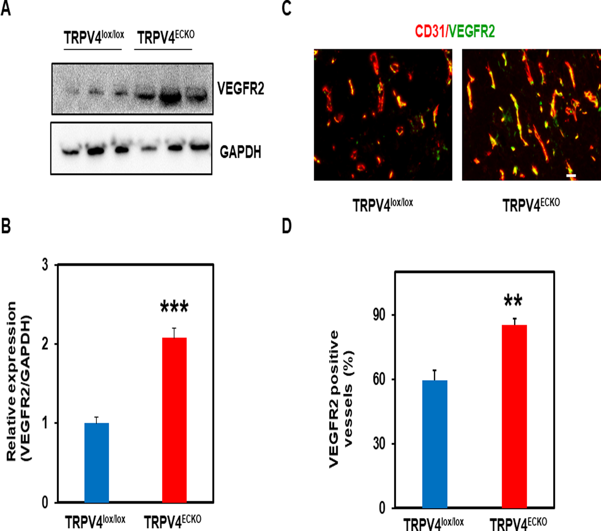
Transient receptor potential vanilloid 4 (TRPV4) is a ubiquitously expressed polymodally activated ion channel. TRPV4 has been implicated in tumor progression; however, the cell-specific role of TRPV4 in tumor growth, angiogenesis, and metastasis is unknown. Here, we generated endothelial-specific TRPV4 knockout (TRPV4ECKO) mice by crossing TRPV4lox/lox mice with Tie2-Cre mice. Tumor growth and metastasis were significantly increased in a syngeneic Lewis lung carcinoma tumor model of TRPV4ECKO mice compared to TRPV4lox/lox mice. Multiphoton microscopy, dextran leakage, and immunohistochemical analysis revealed increased tumor angiogenesis and metastasis that were correlated with aberrant leaky vessels (increased width and reduced pericyte and VE-cadherin coverage). Mechanistically, increases in VEGFR2, p-ERK, and MMP-9 expression and DQ gelatinase activity were observed in the TRPV4ECKO mouse tumors. Our results demonstrated that endothelial TRPV4 is a critical modulator of vascular integrity and tumor angiogenesis and that deletion of TRPV4 promotes tumor angiogenesis, growth, and metastasis.

Keywords: Endothelial cell; Metastasis; Transient receptor potential vanilloid 4; Tumor angiogenesis; Vascular endothelial growth factor receptor 2.


Conflict of interest statement

Conflict of Interest:

The authors declare no conflict of interest.

Figures

Figure 1.
Figure 1.. Characterization of endothelial TRPV4 knockout (TRPV4ECKO) deletion in mice.
A) Immunohistochemistry shows the colocalization (yellow; arrows) of the endothelial marker CD31 (green) with TRPV4 (red; arrowheads) in the TRPV4lox/lox mouse aortae; this finding is absent in TRPV4ECKO mouse aortae (only green). Scale bar: 10 μm. Note that TRPV4 (red) is present only in the smooth muscle layer of the TRPV4ECKO mouse aortae. B) RT-PCR analysis of TRPV4, VEGFR2, and GAPDH expression in isolated mouse aortic endothelial cells from the TRPV4lox/lox and TRPV4ECKO mice. C) Representative average traces showing relative changes in cytosolic calcium in response to GSK1016790A (GSK101) in the Fluo-4/AM-loaded TRPV4lox/lox and TRPV4ECKO endothelial cells (n=64). The arrow represents the time when the cells were stimulated with GSK101 (F/F0 = ratio of normalized Fluo-4 fluorescence intensity relative to time). D) Quantitative analysis of cytosolic calcium influx induced by GSK101 in the TRPV4lox/lox and TRPV4ECKO endothelial cells. All data represented are the mean ± SEM from at least three independent experiments. Statistical significance is indicated by *** p≤0.001.
Figure 2.
Figure 2.. Deletion of endothelial TRPV4 channels promotes tumor growth and abnormal vasculature.
A) Tumor growth curves of the TRPV4lox/lox (n =9) and TRPV4ECKO (n =11) mice bearing Lewis lung carcinoma (LLC) cells. B) Tumor volumes of the TRPV4lox/lox and TRPV4ECKO mice at day 21. C-D) Multiphoton microscopy images of tumor vasculature from the TRPV4lox/lox and TRPV4ECKO mice (n=3) showing the blood vessel diameter. Mice were infused with Alexa Fluor 594-conjugated isolectin B4 prior to tumor extraction. Scale bar: 10 μm. E) Representative immunofluorescence images of tumor sections of the TRPV4lox/lox and TRPV4ECKO mice stained with an endothelial marker (CD31, red) (n=3). Scale bar: 10 μm. F) Quantification of vessel density from sections of the TRPV4lox/lox and TRPV4ECKO mouse tumors. All data are presented as the mean ± SEM, and statistical significance is indicated by * p≤0.05, **p≤0.01, and *** p≤0.001.
Figure 3.
Figure 3.. Endothelial TRPV4 channel deletion disrupts vascular integrity, leading to leaky vasculature.
A) Representative merged immunofluorescence images from tumor sections of the TRPV4lox/lox and TRPV4ECKO mice stained with an endothelial marker (CD31, red) and pericyte marker (NG2, green) (n=3). Scale bar: 10 μm. Quantification of pericyte coverage (NG2, pericytes in green/CD31, EC in red). B) Representative merged immunofluorescence images from the TRPV4lox/lox and TRPV4ECKO mouse tumor sections stained with an endothelial marker (CD31, red) and endothelial adherens junctional protein (VE-cadherin, green) (n=3). Scale bar: 10 μm. Quantification of VE-cadherin coverage (VE-cadherin, adherens junctional protein in green/CD31, ECs in red). C) Representative immunofluorescence images showing the leakage of Texas red dextran from the vasculature of the TRPV4lox/lox and TRPV4ECKO mouse tumors at day 21. Quantification of mean fluorescence intensity (MFI) (n=3). All data are presented as the mean ± SEM, and statistical significance is indicated by **p≤0.01 and *** p≤0.001.
Figure 4.
Figure 4.. Loss of endothelial TRPV4 increases VEGFR2 expression in LLC tumors.
Western blot analysis (A) and quantification (B) of VEGFR2 expression in LLC tumors from the TRPV4lox/lox and TRPV4ECKO mice (n=3). C) Representative merged immunofluorescence images of tumor sections stained with endothelial markers (CD31, red) and VEGFR2 (green) from the TRPV4lox/lox and TRPV4ECKO mice. Scale bar: 10 μm. D) Quantification of VEGFR2-positive vessels in tumor sections from the TRPV4lox/lox and TRPV4ECKO mice (n=3). All data are presented as the mean ± SEM, and statistical significance is indicated by **p≤0.01 and ***p≤0.001.
Figure 5.
Figure 5.. TRPV4 deletion in the endothelium promotes lung metastasis via increased MMP-9 expression in tumors.
Representative H&E-stained images of lung sections (A) and quantification of metastatic foci (B) from the LLC tumor-bearing TRPV4lox/lox (n=9) and TRPV4ECKO mice (n=11). Scale bar: 200 μm. qPCR analysis of relative MMP-9 (C) and MMP-2 (D) expression in the TRPV4lox/lox and TRPV4ECKO mouse tumors (n=3). All data are presented as the mean ± SEM, and statistical significance is indicated by *p≤0.05 and ** p≤0.01, NS: nonsignificant.

References

    1. Adapala RK, Thoppil RJ, Ghosh K, Cappelli HC, Dudley AC, Paruchuri S, Keshamouni V, Klagsbrun M, Meszaros JG, Chilian WM, Ingber DE, Thodeti CK (2016) Activation of mechanosensitive ion channel TRPV4 normalizes tumor vasculature and improves cancer therapy. Oncogene 35 (3):314–322. doi:10.1038/onc.2015.83 - DOI - PMC - PubMed
    1. Adapala RK, Thoppil RJ, Luther DJ, Paruchuri S, Meszaros JG, Chilian WM, Thodeti CK (2013) TRPV4 channels mediate cardiac fibroblast differentiation by integrating mechanical and soluble signals. Journal of molecular and cellular cardiology 54:45–52. doi:10.1016/j.yjmcc.2012.10.016 - DOI - PMC - PubMed
    1. Cappelli HC, Kanugula AK, Adapala RK, Amin V, Sharma P, Midha P, Paruchuri S, Thodeti CK (2019) Mechanosensitive TRPV4 channels stabilize VE-cadherin junctions to regulate tumor vascular integrity and metastasis. Cancer letters 442:15–20. doi:10.1016/j.canlet.2018.07.042 - DOI - PMC - PubMed
    1. Hartmannsgruber V, Heyken WT, Kacik M, Kaistha A, Grgic I, Harteneck C, Liedtke W, Hoyer J, Kohler R (2007) Arterial response to shear stress critically depends on endothelial TRPV4 expression. PLoS One 2 (9):e827. - PMC - PubMed
    1. Liedtke W (2005) TRPV4 plays an evolutionary conserved role in the transduction of osmotic and mechanical stimuli in live animals. J Physiol 567 (Pt 1):53–58 - PMC - PubMed

Publication types

MeSH terms

LinkOut - more resources