. 2021 Mar 3;17(3):e1008979.
doi: 10.1371/journal.ppat.1008979. eCollection 2021 Mar.

Plxdc family members are novel receptors for the rhesus monkey rhadinovirus (RRV)

Affiliations

Plxdc family members are novel receptors for the rhesus monkey rhadinovirus (RRV)

Anna K Großkopf et al. PLoS Pathog. .

Abstract

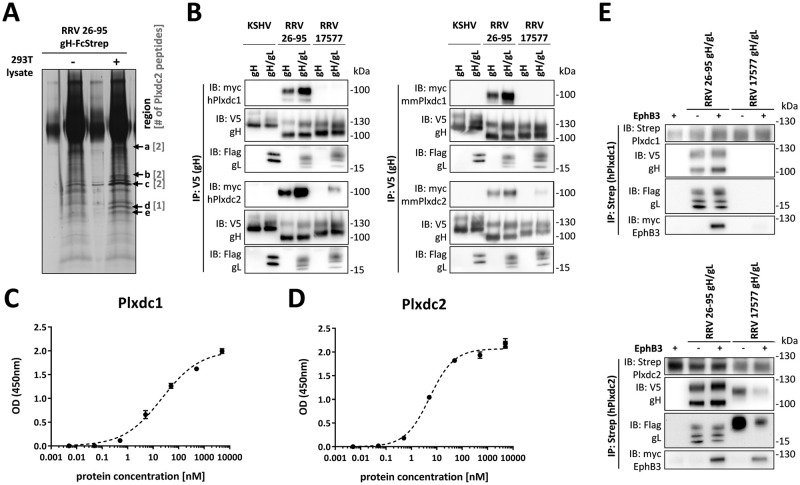
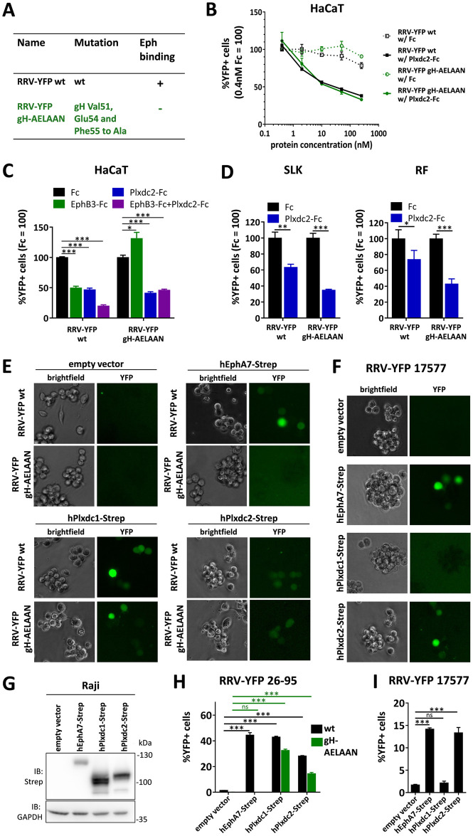
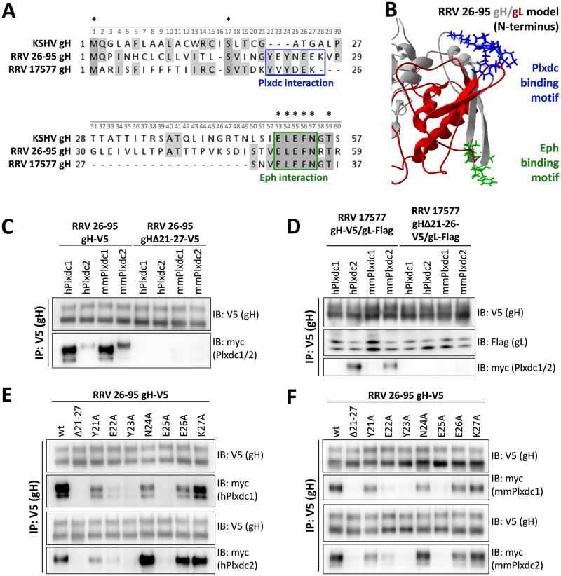
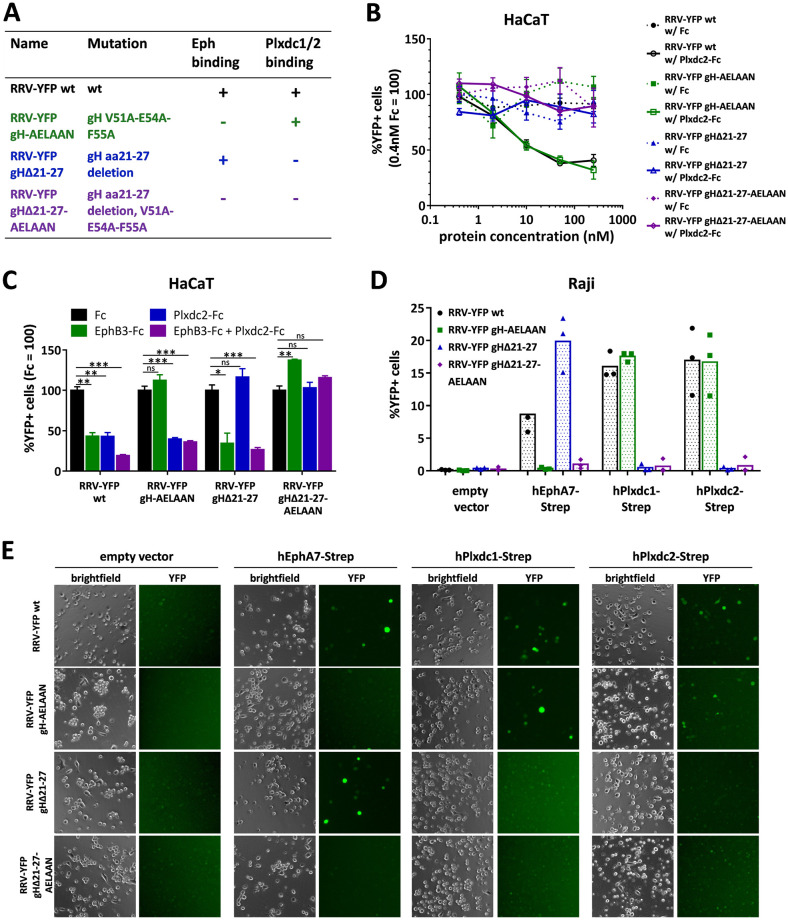
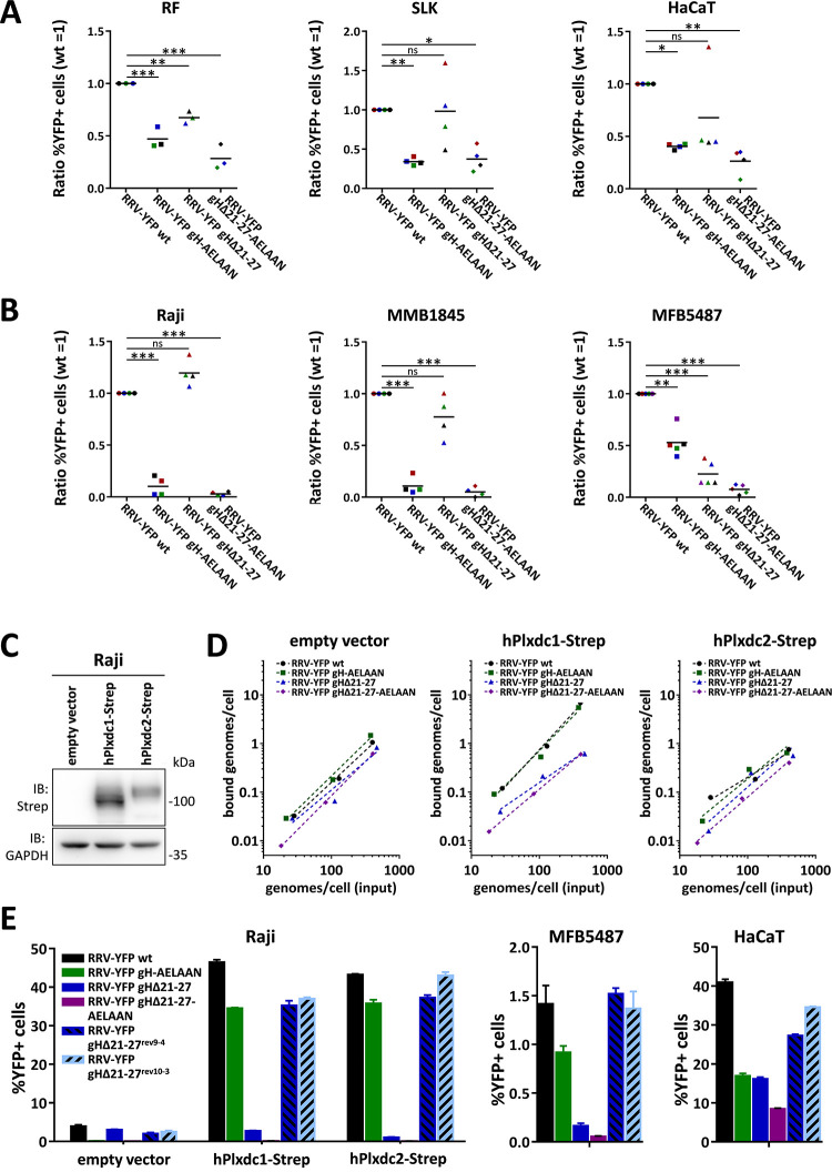
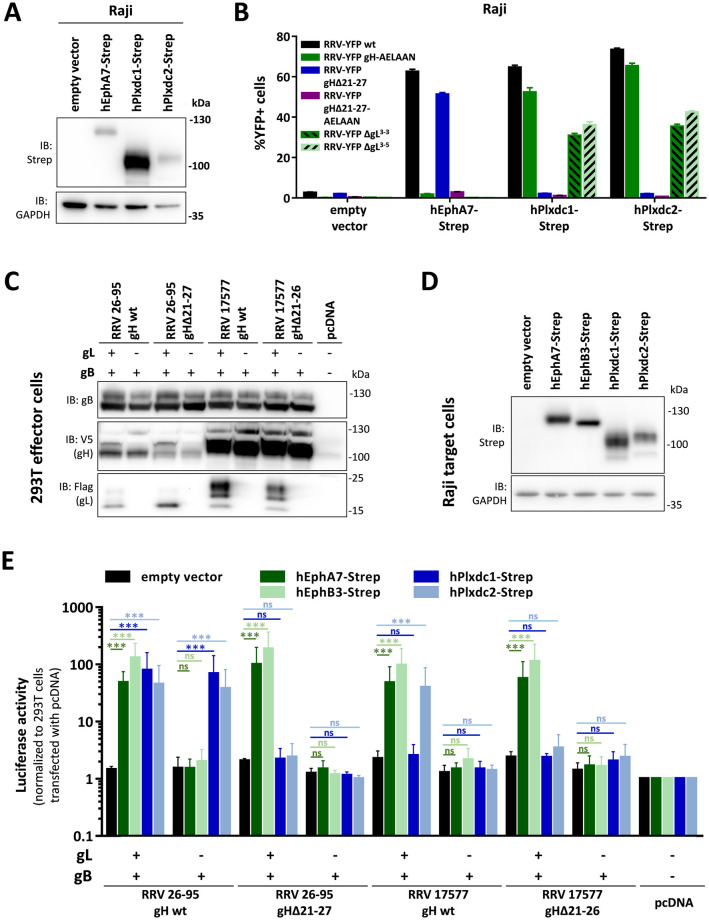
The rhesus monkey rhadinovirus (RRV), a γ2-herpesvirus of rhesus macaques, shares many biological features with the human pathogenic Kaposi's sarcoma-associated herpesvirus (KSHV). Both viruses, as well as the more distantly related Epstein-Barr virus, engage cellular receptors from the Eph family of receptor tyrosine kinases (Ephs). However, the importance of the Eph interaction for RRV entry varies between cell types suggesting the existence of Eph-independent entry pathways. We therefore aimed to identify additional cellular receptors for RRV by affinity enrichment and mass spectrometry. We identified an additional receptor family, the Plexin domain containing proteins 1 and 2 (Plxdc1/2) that bind the RRV gH/gL glycoprotein complex. Preincubation of RRV with soluble Plxdc2 decoy receptor reduced infection by ~60%, while overexpression of Plxdc1 and 2 dramatically enhanced RRV susceptibility and cell-cell fusion of otherwise marginally permissive Raji cells. While the Plxdc2 interaction is conserved between two RRV strains, 26-95 and 17577, Plxdc1 specifically interacts with RRV 26-95 gH. The Plxdc interaction is mediated by a short motif at the N-terminus of RRV gH that is partially conserved between isolate 26-95 and isolate 17577, but absent in KSHV gH. Mutation of this motif abrogated the interaction with Plxdc1/2 and reduced RRV infection in a cell type-specific manner. Taken together, our findings characterize Plxdc1/2 as novel interaction partners and entry receptors for RRV and support the concept of the N-terminal domain of the gammaherpesviral gH/gL complex as a multifunctional receptor-binding domain. Further, Plxdc1/2 usage defines an important biological difference between KSHV and RRV.


Conflict of interest statement

The authors have declared that no competing interests exist.

Figures

Fig 1
Fig 1. Plxdc family receptors are novel interaction partners for RRV gH/gL.
A) Immunoprecipitation of recombinant soluble RRV 26–95 gH-FcStrep in the presence or absence of 293T lysate. Precipitates were analyzed by PAGE, silver stained and bands at the indicated molecular weight (arrows, regions a-e) were excised and analyzed by mass spectrometry. Numbers in brackets indicate the number of Plxdc2 peptides identified by LC-MS/MS in each region. B) V5-tagged RRV 26–95 gH, RRV 17577 gH or KSHV gH alone or co-expressed with the respective Flag-tagged gL construct were immunoprecipitated in the presence of full-length Plxdc1 or Plxdc2 of human (h) or Macaca mulatta (mm) origin using monoclonal antibody to the V5-tag. Precipitates were analyzed by Western blot with the indicated antibodies. C-D) Binding of Plxdc1 ectodomain C) or Plxdc2 ectodomain D) at various concentrations to immobilized RRV 26–95 gH-FcStrep/gL was measured by enzyme-linked immunosorbent assay. Curve Fitting and determination of half-maximal binding concentration was performed based on the One site specific binding with Hill Slope equation in Prism6. E) Co-immunoprecipitation of soluble human Plxdc1-FcStrep or human Plxdc2-FcStrep with RRV 26–95 gH-V5/gL-Flag or RRV 17577 gH-V5/gL-Flag using StrepTactin sepharose in the presence or absence of human full-length myc-tagged EphB3. Abbreviations: IP: immunoprecipitation, IB: immunoblotting, h: human, mm: Macaca mulatta (rhesus macaque).
Fig 2
Fig 2. Plxdc1/2 function as entry receptors for RRV.
A) List of BAC-derived recombinant viruses and introduced mutations used in this figure. B) Dose-dependent inhibition of RRV 26–95 infection by soluble human Plxdc2-FcStrep on HaCaT cells. RRV-YFP wt and RRV-YFP gH-AELAAN were pre-incubated with hPlxdc2-FcStrep for 30min at room temperature. FcStrep alone was used as control. YFP expression as indicator of infection was measured by flow cytometry. Infection in the presence of 0.4nM FcStrep was set to 100% (MOI ~0.2, triplicates, error bars represent SD). C) Inhibition of RRV 26–95 infection by soluble Plxdc2-FcStrep and EphB3-Fc on HaCaT cells. RRV-YFP wt and RRV-YFP gH-AELAAN were pre-incubated with 100nM hPlxdc2-FcStrep, 10nM EphB3-Fc or a combination of 100nM hPlxdc2-FcStrep and 10nM EphB3-Fc for 30min at room temperature. FcStrep alone was used as control. YFP expression as indicator of infection was measured by flow cytometry. Infection with FcStrep was set to 100% (MOI ~0.2, triplicates, error bars represent SD). D) Inhibition of RRV 26–95 infection by soluble human Plxdc2-FcStrep on SLK cells and rhesus monkey fibroblasts (RF). RRV-YFP wt and RRV-YFP gH-AELAAN were pre-incubated with 250nM hPlxdc2-FcStrep for 30min at room temperature. FcStrep alone was used as control. YFP expression as indicator of infection was measured by flow cytometry. Infection with FcStrep was set to 100% (MOI ~0.05–0.1, triplicates, error bars represent SD). E-F) Raji cells were transduced with TwinStrep-tagged human Plxdc1, Plxdc2 or EphA7 (hPlxdc1-Strep/ hPlxdc2-Strep/ hEphA7-Strep) expression constructs or an empty vector control, briefly selected and infected with RRV-YFP 26–95 wt and RRV-YFP 26–95 gH-AELAAN (E) or RRV-YFP 17577 (F) normalized to genome copies as determined by qPCR. Micrographs show representative infection of the indicated cell pools. G) Lysates of transduced Raji cell pools were analyzed for EphA7-Strep and Plxdc1/2-Strep expression by Western blot. H) Quantification of (E) by flow cytometric analyses of YFP reporter gene expression as indicator of infection (triplicates, error bars represent SD). I) Quantification of (F) by flow cytometric analyses of YFP reporter gene expression as indicator of infection (triplicates, error bars represent SD).
Fig 3
Fig 3. An amino acid sequence motif in the N-terminal region of RRV 26–95 and RRV 17577 gH is essential for the Plxdc interaction.
A) Multiple sequence alignment of the N-terminal region of gH of KSHV and the two RRV isolates 26–95 and 17577. Boxes indicate the binding motives for Eph receptors (green) and Plxdc receptors (blue). Numbers above sequences represent positions in the alignment, asterisks indicate conserved amino acids. Individual amino acid positions are given to the left and right of the respective gH sequence. B) Homology-based structure prediction of the RRV 26–95 gH/gL complex based on the crystal structure of the EBV gH/gL complex (PDB number 3PHF) using the Iterative Threading ASSembly Refinement (I-TASSER) server and the CO-THreader (COTH) algorithms for protein-protein complex structure and multi-chain protein threading. The Eph receptor interaction motif is shown in green, the Plxdc interaction motif is shown in blue, gL is shown in red, gH is shown in grey. C) Deletion of the Plxdc interaction motif in RRV 26–95 gH (amino acid 21–27, “YEYNEEK”) abrogates gH interaction with Plxdc1 and Plxdc2. V5-tagged gH wt or gHΔ21–27 were immunoprecipitated in the presence of full-length human or Macaca mulatta Plxdc1-myc or Plxdc2-myc using monoclonal antibody to the V5-tag. Precipitates were analyzed by Western blot. D) Deletion of the Plxdc interaction motif in RRV 17577 gH (amino acid 21–26, “YVYDEK”) abrogates gH interaction with Plxdc2. V5-tagged gH wt or gHΔ21–26 was co-expressed with Flag-tagged RRV 17577 gL. gH-V5/gL-Flag complexes were immunoprecipitated in the presence of full-length human or Macaca mulatta Plxdc1-myc or Plxdc2-myc using monoclonal antibody to the V5-tag. Precipitates were analyzed by Western blot. E) Mutational scan of the Plxdc interaction motif (amino acid 21–27, “YEYNEEK”) of RRV 26–95 gH identifies human Plxdc1/2-interacting residues. V5-tagged gH mutants were immunoprecipitated in the presence of full-length human Plxdc1-myc or Plxdc2-myc using monoclonal antibody to the V5-tag. Precipitates were analyzed by Western blot. RRV gHΔ21–27 serves as negative control. F) Mutational scan of the Plxdc interaction motif (amino acid 21–27, “YEYNEEK”) of RRV 26–95 gH identifies rhesus macaque Plxdc1/2-interacting residues. V5-tagged gH mutants were immunoprecipitated in the presence of full-length Macaca mulatta Plxdc1-myc or Plxdc2-myc using monoclonal antibody to the V5-tag. Precipitates were analyzed by Western blot. RRV gHΔ21–27 serves as negative control. Abbreviations: IP: immunoprecipitation, IB: immunoblotting, h: human, mm: Macaca mulatta (rhesus macaque).
Fig 4
Fig 4. Deletion of the seven amino acid Plxdc-binding motif is sufficient to detarget RRV 26–95 from Plxdc receptors.
A) List of BAC-derived recombinant viruses and introduced mutations used in this figure. B) Dose-dependent inhibition of RRV 26–95 infection by soluble human Plxdc2-FcStrep on HaCaT cells. RRV-YFP wt, RRV-YFP gH-AELAAN, RRV-YFP gHΔ21–27 and RRV-YFP gHΔ21-27-AELAAN were pre-incubated with hPlxdc2-FcStrep for 30min at room temperature. FcStrep alone was used as control. YFP expression as indicator of infection was measured by flow cytometry. Infection in the presence of 0.4nM FcStrep was set to 100% (MOI ~0.05, triplicates, error bars represent SD). C) Inhibition of RRV 26–95 infection by soluble Plxdc2-FcStrep and EphB3-Fc on HaCaT cells. RRV-YFP wt, RRV-YFP gH-AELAAN, RRV-YFP gHΔ21–27 and RRV-YFP gHΔ21-27-AELAAN were pre-incubated with 100nM hPlxdc2-FcStrep, 10nM EphB3-Fc or a combination of 100nM hPlxdc2-FcStrep and 10nM EphB3-Fc for 30min at room temperature. FcStrep alone was used as control. YFP expression as indicator of infection was measured by flow cytometry. Infection with FcStrep was set to 100% (MOI ~0.1–0.2, triplicates, error bars represent SD). D) Raji cells were transduced with TwinStrep-tagged human EphA7, Plxdc1 or Plxdc2 (hEphA7-Strep/ hPlxdc1-Strep/ hPlxdc2-Strep) expression constructs or an empty vector control, briefly selected and infected with RRV-YFP wt, RRV-YFP gH-AELAAN, RRV-YFP gHΔ21–27 or RRV-YFP gHΔ21-27-AELAAN normalized to genome copies as determined by qPCR. YFP expression as indicator of infection was measured by flow cytometry. The mean across three independent sets of RRV stocks is indicated by columns. The means of individual triplicate infections for each set of RRV stocks are given as symbols within the respective columns. E) Micrographs show representative infection of one set of RRV stocks in (D).
Fig 5
Fig 5. The contribution of the RRV 26–95 gH-Plxdc interaction to infection is cell type-specific and may in part be dependent on attachment effects.
A) RRV 26–95 deleted in the Plxdc interaction motif exhibits reduced specific infectivity on HaCaT and RF, but not SLK cells. Target cells were infected with RRV-YFP wt, RRV-YFP gH-AELAAN, RRV-YFP gHΔ21–27 or RRV-YFP gHΔ21-27-AELAAN normalized to genome copies as determined by qPCR. YFP expression as indicator of infection was measured by flow cytometry and normalized to RRV-YFP wt infection. Means of individual normalized infections with three (RF) or four (HaCaT, SLK) independent sets of RRV stocks are given as symbols of different color. Sets with RRV-YFP wt infection in an MOI range of 0.05–1 were used for analysis. The mean across the independent sets of RRV stocks is indicated by black lines. B) RRV 26–95 deleted in the Plxdc interaction motif exhibits reduced specific infectivity on MFB5487, but not Raji and MMB1845 cells. Target cells were infected with RRV-YFP wt, RRV-YFP gH-AELAAN, RRV-YFP gHΔ21–27 or RRV-YFP gHΔ21-27-AELAAN normalized to genome copies as determined by qPCR. YFP expression as indicator of infection was measured by flow cytometry and normalized to RRV-YFP wt infection. Means of individual normalized infections with four (Raji, MMB1845) or five (MFB5487) independent sets of RRV stocks are given as symbols of different color. Sets with RRV-YFP wt infection exceeding 1% were used for analysis. The maximal achieved RRV-YFP wt infection was 6.1% for Raji, 5.6% for MMB1845 and 3.1% for MFB5487, respectively. The mean across the independent sets of RRV stocks is indicated by black lines. C) Western blot analysis of Raji cells transduced with TwinStrep-tagged human Plxdc1 and Plxdc2 (hPlxdc1-Strep/ hPlxdc2-Strep) expression constructs or an empty vector control. D) Attachment of RRV 26–95 on transduced Raji cells is affected by hPlxdc1-Strep, but not hPlxdc2-Strep overexpression. Cells, analyzed in (C), were incubated with cold virus at the indicated concentrations at 4°C for 30min followed by genomic DNA isolation. Bound genomes/cells as calculated based on qPCR of a genomic (CCR5) and a viral locus (ORF73/ LANA) were plotted against input viral genome number determined by ORF73/ LANA qPCR. E) Re-introduction of the seven amino acid motif crucial for Plxdc interaction rescues RRV-YFP gHΔ21–27 infection. Transduced Raji cells, MFB5487 and HaCaT cells were infected with RRV-YFP wt, RRV-YFP gH-AELAAN, RRV-YFP gHΔ21–27, RRV-YFP gHΔ21-27-AELAAN or two RRV-YFP gHΔ21–27 revertants (RRV-YFP gHΔ21-27rev9-4, RRV-YFPgHΔ21-27rev10-3) normalized to genome copies as determined by qPCR. YFP expression as indicator of infection was measured by flow cytometry (triplicates, error bars represent SD).
Fig 6
Fig 6. Plxdc1/2-dependent infection with RRV 26–95 does not require gL.
A) Raji cells were transduced with TwinStrep-tagged human EphA7, Plxdc1 and Plxdc2 (hEphA7-Strep/ hPlxdc1-Strep/ hPlxdc2-Strep) expression constructs or an empty vector control and briefly selected by antibiotic resistance. Lysates of transduced Raji cell pools were analyzed for EphA7-Strep and Plxdc1/2-Strep expression by Western blot. B) Transduced Raji cells analyzed in (A) were infected with RRV-YFP wt, RRV-YFP gH-AELAAN, RRV-YFP gHΔ21–27 or one of two RRV-YFP ΔgL clones normalized to genome copies as determined by qPCR. YFP expression as indicator of infection was measured by flow cytometry (triplicates, error bars represent SD). C-E) Cell-cell fusion assay: Transfected 293T effector cells were co-cultured with transduced Raji target cells as indicated. Cell-cell fusion was assessed after two days of co-culture. Protein expression of 293T effector cells transfected with empty vector or the indicated viral glycoprotein combinations (C) or of transduced Raji target cells (D) was analyzed by Western blot. Expression controls were harvested directly prior to the start of co-culture. Luciferase activity as indicator of cell-cell fusion was measured after 48h of effector and target cell co-culture (E). Luciferase activity is normalized to empty vector transfected 293T effector cells (n = 3, error bars represent SD).

Similar articles

Cited by

References

    1. Desrosiers RC, Sasseville VG, Czajak SC, Zhang X, Mansfield KG, Kaur A, et al.. A herpesvirus of rhesus monkeys related to the human Kaposi’s sarcoma-associated herpesvirus. J Virol. 1997;71: 9764–9769. 10.1128/JVI.71.12.9764-9769.1997 - DOI - PMC - PubMed
    1. Searles RP, Bergquam EP, Axthelm MK, Wong SW. Sequence and genomic analysis of a Rhesus macaque rhadinovirus with similarity to Kaposi’s sarcoma-associated herpesvirus/human herpesvirus 8. J Virol. 1999;73: 3040–3053. 10.1128/JVI.73.4.3040-3053.1999 - DOI - PMC - PubMed
    1. O’Connor CM, Kedes DH. Rhesus Monkey Rhadinovirus: A Model for the Study of KSHV. In: Boshoff C, Weiss RA, editors. Kaposi Sarcoma Herpesvirus: New Perspectives. Springer; Berlin Heidelberg; 2007. pp. 43–69. - PubMed
    1. Shin YC, Jones LR, Manrique J, Lauer W, Carville A, Mansfield KG, et al.. Glycoprotein gene sequence variation in rhesus monkey rhadinovirus. Virology. 2010;400: 175–186. 10.1016/j.virol.2010.01.030 - DOI - PMC - PubMed
    1. Bilello JP, Morgan JS, Damania B, Lang SM, Desrosiers RC. A Genetic System for Rhesus Monkey Rhadinovirus: Use of Recombinant Virus To Quantitate Antibody-Mediated Neutralization. J Virol. 2006;80: 1549–1562. 10.1128/JVI.80.3.1549-1562.2006 - DOI - PMC - PubMed

Publication types

MeSH terms