Review
. 2021 May:126:63-91.
doi: 10.1016/j.actbio.2021.02.034. Epub 2021 Feb 28.

Natural medicine delivery from biomedical devices to treat bone disorders: A review

Affiliations
Review

Natural medicine delivery from biomedical devices to treat bone disorders: A review

Susmita Bose et al. Acta Biomater. 2021 May.

Abstract

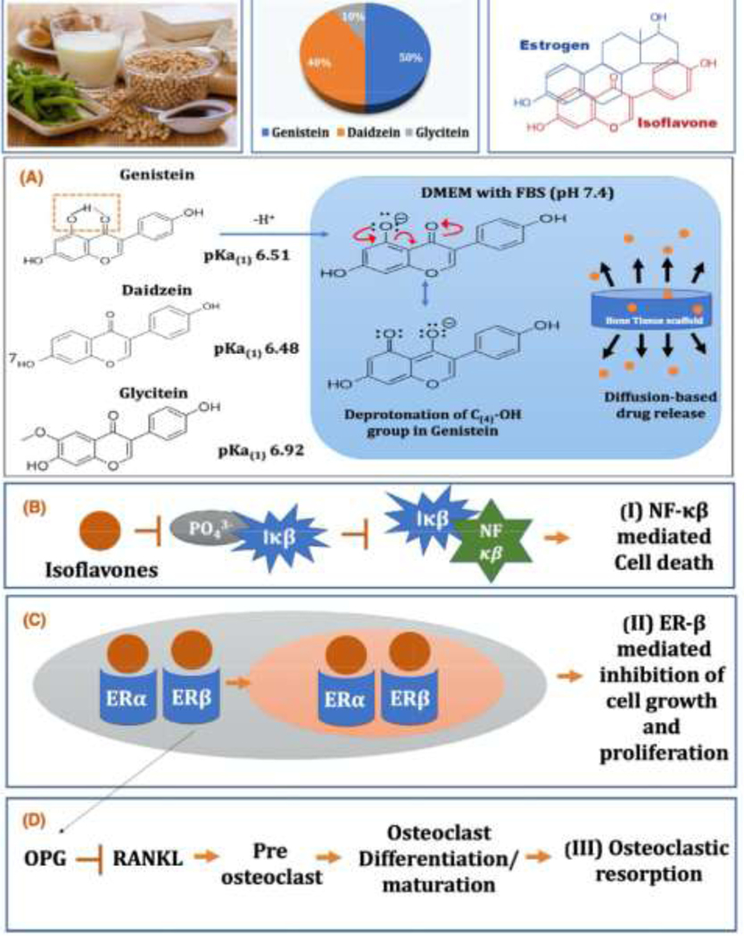
With an increasing life expectancy and aging population, orthopedic defects and bone graft surgeries are increasing in global prevalence. Research to date has advanced the understanding of bone biology and defect repair mechanism, leading to a marked success in the development of synthetic bone substitutes. Yet, the quest for functionalized bone grafts prompted the researchers to find a viable alternative that regulates cellular activity and supports bone regeneration and healing process without causing serious side-effects. Recently, researchers have introduced natural medicinal compounds (NMCs) in bone scaffold that enables them to release at a desirable rate, maintains a sustained release allowing sufficient time for tissue in-growth, and guides bone regeneration process with minimized risk of tissue toxicity. According to World Health Organization (WHO), NMCs are gaining popularity in western countries for the last two decades and are being used by 80% of the population worldwide. Compared to synthetic drugs, NMCs have a broader range of safety window and thus suitable for prolonged localized delivery for bone regeneration. There is limited literature focusing on the integration of bone grafts and natural medicines that provides detailed scientific evidences on NMCs, their toxic limits and particular application in bone tissue engineering, which could guide the researchers to develop functionalized implants for various bone disorders. This review will discuss the emerging trend of NMC delivery from bone grafts, including 3D-printed structures and surface-modified implants, highlighting the significance and potential of NMCs for bone health, guiding future paths toward the development of an ideal bone tissue engineering scaffold. STATEMENT OF SIGNIFICANCE: To date, additive manufacturing technology provids us with many advanced patient specific or defect specific bone constructs exhibiting three-dimensional, well-defined microstructure with interconnected porous networks for defect-repair applications. However, an ideal scaffold should also be able to supply biological signals that actively guide tissue regeneration while simultaneously preventing post-implantation complications. Natural biomolecules are gaining popularity in tissue engineering since they possess a safer, effective approach compared to synthetic drugs. The integration of bone scaffolds and natural biomolecules exploits the advantages of customized, multi-functional bone implants to provide localized delivery of biochemical signals in a controlled manner. This review presents an overview of bone scaffolds as delivery systems for natural biomolecules, which may provide prominent advancement in bone development and improve defect-healing caused by various musculoskeletal disorders.

Keywords: Biomaterials; Bone disorders; Bone tissue engineering; Drug delivery; Natural medicinal compounds; Vitamins.


Conflict of interest statement

Declaration of Competing Interest The authors declare that they have no known competing financial interests or personal relationships that could have appeared to influence the work reported in this paper.

Figures

Fig.1
Fig.1
Integration of natural medicinal compounds and bone grafts: Localized delivery of natural medicinal compounds from bone scaffolds can be a promising alternative to conventional bone grafts that provides controlled delivery of biochemical cues to the target sites without inducing adverse effects on healthy bone. (A) Biological sources of various natural medicinal compounds (B) bone cement injection in vertebroplasty (C) Evolution of dental implants over the years, from titanium implant with zirconia top, one-piece zirconia implant to current two-piece ceramic implants. (D) synthetic 3D printed cranial implants, patient specific jaw reconstruction c) [13] (E) Cemented joint prosthesis, that uses a quick-setting bone cement that bonds with patient’s natural bone. Cementless or press-fit implants can be coated with HA which enhance bone growth onto the porous coating surface (F) (i) The 3D pelvis was segmented by the thresholding process in the CAD software. The extent of tumor was outlined on each axial CT image and its tumor volume was extracted (red in color). A 3D bone tumor model was created for the surgical planning. (ii) Surgeons performed the virtual resections by defining the locations and orientations of the resection planes. (iii) The virtually resected tumor was extracted. As the bone was deformed by the tumor, the mirrored 3D image from the normal side of the hemipelvis was used to duplicate the core shape of the implant. (iv) The flanges and the acetabular cup were added for better implant stability. The final implant design had a normal acetabular contour and the components for implant fixation. [14].
Fig. 2
Fig. 2
Natural medicinal compounds for the treatment of various musculoskeletal disorders: Natural medicines with distinct biological activities can be optimized for effective dosage concentration and desired release kinetics. Then it can be incorporated within tissue engineering grafts to repair bone defects caused by surgical intervention due to various musculoskeletal disorders including osteoporosis, osteosarcoma, osteomyelitis, osteoarthritis, and rheumatoid arthritis. Localized delivery of natural medicines in an optimized dosage might promote bone defect repair, lower the dosage frequency of systemic drug administration and minimize tissue toxicity caused by high dosages of synthetic drugs.
Fig. 3
Fig. 3
Bioavailability, controlled release kinetics and tissue materials interaction are three important criteria for a clinically successful localized drug delivery system. Poor bioavailability of naturally sourced biomolecules often creates a barrier in their practical application. In such cases, various drug delivery carriers have been employed to control the release kinetics and subsequently enhance the bioavailability. A controlled release of biomolecules from bone grafts at a desired concentration improves host tissue and implant materials interaction and provides biological functionality to the scaffold for treating different musculoskeletal disorders.
Fig. 4
Fig. 4
Efficacy of acemannan in bone formation in vitro and in vivo. (A) Acemannan significantly enhanced periodontal ligament cell ALPase activity after 72 h of incubation at concentrations of 2 and 4 mg/mL. [*denotes statistical difference with the untreated group; P < 0.05, n = 3] (B-C) Acemannan increased periodontal ligament cell mineral deposition at 9 and 18 day. The 0.5, 1, 2 and 4 mg/mL acemannan-treated groups had significantly higher mineralization as shown by more intensely stained areas by alizarin-red staining compared to the untreated group [*, # Compared with the untreated group at the 9th and 18th day of incubation, respectively, P < 0.05, n = 9] (D) Enhanced bone mineral density (BMD) was also noted in a dog tooth model 4 weeks after extraction[*, # Significant difference compared to the untreated socket; p < 0.05, n = 7] (E). Histopathology of the tooth socket demonstrates numerous thick and dense bone trabeculae bridges in the acemannan treated group compared to large unfilled zones in the control group after Hematoxylin and Eosin (H&E) staining bar 200 μm (a1, b1) and 50 μm (a2, b2) [–52].
Fig. 5
Fig. 5
Possible mechanism of action of acemannan-mediated bone remodeling. Acemannan stimulates the production of cytokines IL-6 and TNF- α:, which allows pre-osteoblasts to form osteoblast and express RANKL. Osteoblast-derived RANKL and M-CSF bind to RANK and c-FMS receptor on monocytes and differentiate them to mature osteoclasts. [IL-6: Interleukin-6, TNF- α: tumor necrosis factor-alpha, RANK: receptor activator of nuclear factor kappa B, RANKL: receptor activator of nuclear factor kappa B ligand, M-CSF: macrophage colony stimulating factor, c-FMS: Colony-stimulating Factor-1 Receptor. [Adapted from 53].
Fig. 6
Fig. 6
The efficacy of acemannan released from HA coated Ti implants on in vitro osteoblast cells and in vivo bone regeneration in rat distal femur model. (A) Aloe vera gel and it’s chief active component, acemannan. (B) After 1 day of incubation, osteoblast cell density increased significantly in presence of acemannan compared to HA coating. However, the cell viability decreased when acemannan concentration was more than 500 μg/ml of FBS enriched DMEM. (C) Controlled release of acemannan from TCP matrix has been achieved in physiological pH for 14 days. (D) CT scan and SEM micrographs of HA-coated Ti64 implants shows the presence of acemannan improved the osteoid formation, evident from the enhanced reddish orange color around the vicinity of the implant. SEM micrographs also showed no gaps at the interface of the acemannan implants. The combined presence of chitosan with acemannan further mineralized the new osteoid formation around the implant, shown by the greenish blue color, leading to improved early stage osseointegration. Osseous tissue had been observed to interlock with the implant in the SEM micrographs, leaving no visible gaps. (E) Histomorphometric assay performed using the ImageJ software from the histology micrographs of Ti6Al4V implants. Presence of acemannan significantly improved the osteoid formation. [54].
Fig.7
Fig.7
Mechanism of action of curcumin via NFκB pathway shows curcumin blocks phosphorylation and degradation of NF–κB inhibitor (IκB), which leads to formation of inactive NF–κB-IκB conjugate. Suppression of NF-κB positively influence osteoblast differentiation and prevents osteoclastic bone resorption [Adapted from 58–60].
Fig.8
Fig.8
(A) Effects of curcumin on the bone metabolism of rats with dexamethasone (DXM)-induced osteoporosis. Rats with DXM-induced osteoporosis exhibited significantly decreased osteocalcin levels and increased collagen-type I fragments (CTX) serum levels. The administration of curcumin reversed these changes [##P<0.01 compared to the control group, **P<0.01 compared to the DXM group]. (B) Effects of curcumin on the Wnt signaling pathway in primary osteoblasts. (top) The mRNA expression levels of Wnt, β-catenin, low-density lipoprotein receptor-related protein 5 (LRP5), sclerostin (SOST) and Dickkopf-1 in DXM-stimulated primary osteoblasts [##P<0.01 compared to the control group, **P<0.01 compared to the DXM group]. (middle) The phosphorylation of glycogen synthase kinase-3β (GSK-3β) in dexamethasone (DXM)-stimulated primary osteoblasts. β-catenin trafficking in DXM-stimulated primary osteoblasts. (bottom) DXM significantly inhibited the Wnt signaling pathway, and curcumin prevented the inhibition (Scale bar, 50 μm). (C) Histological study using H&E staining in rat femur showing effects of curcumin in dexamethasone (DXM)-induced osteoporosis. DXM caused obvious damage to the femurs of rats and curcumin attenuated the damage (Scale bar, 100 μm) [63].
Fig. 9
Fig. 9
Effect of curcumin released from calcium phosphate matrix for osteoblast in vitro proliferation and in vivo bone regeneration (a) Osteoblast (hFOB) cell viability of after day 3,7 and 11 days of culture by MTT assay (n=9). Curcumin with PCL-PEG showing statistically significant difference compared to the untreated TCP control. [*** denotes P value < 0.0001 compared to control, “ns” denotes not statistically significant compared to control]. (b) SEM images for osteoblast cell culture of control, control+curcumin+PCL/PEG samples after 7 days of culture. Regardless of the time point, polymeric curcumin coated samples exhibiting much higher cell proliferation compared to samples without polymer (c) Optical microscopy images of tissue-implant sections after von Willebrand Factor (vWF) and modified Masson Goldner trichrome staining showing angiogenesis and osteoid like new bone formation respectively after 6 weeks of surgery in rat distal femur model (d) Histomorphometric analysis using ImageJ showing enhanced osteoid like new bone formation in curcumin coated TCP scaffolds compared to control TCP scaffolds after 6 weeks of surgery. [*** denote P values < 0.0001 compared to control] [70].
Fig.10
Fig.10
The potency of gingerol extract (0–50 mM) on collagen production and ALP mRNA expression with and without stimulation of tumor necrosis factor (TNF)-α. Data are reported as mean±SE for 3 independent experiments. #P,0.05 compared to 0 mM; *P,0.05 compared to control (Con) (Student’s t-test). Results suggest that gingerol enhances TNF-α suppressed osteoblast like cell activity and differentiation, revealing beneficial effects of gingerol on treatment of bone disorders [97].
Fig.11
Fig.11
Mechanism of action of vitamin B6, vitamin B9 (Folic Acid) and vitamin B12 in reducing osteoclast activity. Vitamin B6, B9 and B12 directly influence homocysteine metabolism. Upregulation of homocysteine can be correlated to the reactive oxygen species (ROS) formation and NF-κB activation, and subsequent osteoclastic activity [Adapted from 107].
Fig.12
Fig.12
The graphical presentation shows the dietary sources of soy isoflavones, percentage constituents of isoflavones found in soy foods and similarity of isoflavones to mammalian estrogen. The chemical structure of primary three isoflavones, genistein, daidzein and glycitein are also presented. (A) The acid dissociation constant or pKa values for genistein, daidzein and glycitein are 6.51, 6.48 and 6.92, respectively, indicating that these isoflavones are easily deprotonated at physiological pH of 7.4, resulting in overall higher resonance stability and solubility in physiological pH (B) The possible mechanism of action of soy isoflavones towards chemoprevention and anti-inflammatory activity is demonstrated through NFκB inhibition pathway. (C) Estrogen-mimicking activity of soy isoflavones result in its ability to bind to the estrogen receptor and subsequent ER-β-mediated inhibition of cell growth and proliferation. (D) Isoflavones also suppress osteoclastogenesis through the upregulation of OPG expression, which in turn inhibits RANK-RANKL interactions [130].
Fig.13
Fig.13
(A) FESEM images showing the effects of soy isoflavones loaded 3DP tri calcium phosphate (TCP) bone tissue engineering scaffolds for in vitro osteoblast cell proliferation in a flow perfusion bioreactor at day 5. Combination of soy isoflavones, genistein, daidzein and glycitein in 5:4:1 ratio show presence of higher osteoblast proliferation compared to control. Interestingly, more cells could be seen near the designed pores of the 3DP scaffolds indicating the importance of flow transport through the porous channel. (B) MTT assay (n = 3) showing the effects of soy isoflavones loaded 3DP TCP bone tissue engineering scaffolds for in vitro osteoblast cell proliferation in a flow perfusion bioreactor at day 5 and 10.) [** denotes P-value <0.0001, statistically significant difference between control and test sample]. [Control: 3D TCP: 3D printed porous TCP scaffold] (C) ALP assay (n = 3) showing the effects of soy isoflavones loaded 3DP TCP bone tissue engineering scaffolds for in vitro osteoblast cell differentiation in a flow perfusion bioreactor at day 10. [** denotes P-value <0.0001, statistically significant difference between control and test sample]. [Control: 3D TCP: 3D printed porous TCP scaffold]. (D) Optical microscopy images of decalcified tissue-implant specimens after H&E staining showing inflammatory cell recruitment after 24 hours of surgery in rat distal femur model. Blue and black arrow show neutrophil recruitment and presence of osteocytes, respectively. (E) FESEM images showing the effects of soy isoflavones loaded 3DP TCP bone tissue engineering scaffolds for in vitro MG-63 cell proliferation in a static condition at day 3, 7 and 11. Layers of bone cancer cells can be seen on TCP scaffold alone, where the presence of genistein exhibits lower cell proliferation suggesting its chemopreventive ability. Although daidzein and glycitein did not have a pronounced effect on MG-63 cells on day 7, the combined effect of three isoflavones showed significant chemopreventive effects in all the time points. [131].
Fig.14
Fig.14
(A) Dietary sources of vitamin K and its mechanism of action: vitamin K is essential for the conversation of glutamate to Gla protein, an important constituent of osteocalcin, a bone protein. Presence of carboxylated osteocalcin in blood indicates bone formation [140]. (B) In vivo Masson Goldner staining by curcumin, vitamin K2, and curcumin + vitamin K2 loaded HA-coated Ti implant showing the gap between the implant and the surrounding bone tissue after 5 days of implantation at rat distal femur model. The new bone formation or osteoid tissue is stained with red/orange; mineralized bone tissue is stained with green/blue, and the implant can be seen in black. (B) Histomorphometric analysis using ImageJ software showing percentage osteoid or new bone tissue formation and percentage total bone formation within 250 μm radius of implant after 5 days. Drug-loaded implant exhibited statistically significant difference in osteoid formation and total bone formation after 5 days indicating better osseointegration ability compared to the untreated HA coated Ti control (*P < 0.001 compared to control, **P < 0.05 compared to control) [141].
Fig.15
Fig.15
(A) Possible mechanism of action of vitamin C on inhibition of tumor cells and proliferation of osteoblast cells. The oxidized form of vitamin C or dehydroascorbate (DHA) is taken up by the cells via glucose transporters, which is also upregulated in tumor cells compared to normal cells. Therefore, tumor cells automatically uptake more glucose along with DHA compared to normal cells. Inside the cell, DHA is again reduced back to vitamin C, where it gets accumulated and acts as a pro-oxidant that produces oxidative stress. Generation of free radicals or reactive oxygen species (ROS) and hydrogen peroxide (H2O2) causes cellular damage, suppresses tumor growth and subsequently results in vitamin C-mediated cell death [174]. (B) MTT osteosarcoma cell viability assay showing presence of vitamin C has significantly decreased osteosarcoma cell viability by almost 2.5-fold at day 7 [* denotes P < 0.05, statistically significant difference compared to untreated HA coated Ti control]. (C) SEM images of osteosarcoma cell culture on vitamin C loaded HAp coated cpTi samples at day 3 and day 7 showing significantly reduced osteosarcoma cell proliferation in samples with 25 mM vitamin C. (D) Osteoblast cell viability and ALP activity on control and vitamin C loaded scaffolds after 3, 7, and 11 days of culture showing a pronounced increase in osteoblast cell proliferation in the presence of vitamin C compared to control. Data are presented as mean ± standard deviation (* P < 0.05 compared to untreated TCP control). (E) Osteoblast cellular morphology on scaffolds after 7 days of culture showing a clear distinction between vitamin C scaffolds with the control. Layers of osteoblast cells can be seen in scaffolds containing vitamin C compared to control [–178].
Fig.16
Fig.16
(A) Mechanism of action of vitamin D3 (VD3) mediated bone formation and mineralization and inhibition of osteoclast activity [191]. (B) Osteoclast resorption pit assay showing clear resorption pits on HA and HA loaded with polycaprolactone/ polyethylene glycol (PCL/PEG) samples however little to no resorption pits could be found on samples loaded with VD3 indicating reduced osteoclast activity. (C) SEM images with hFOB after 7 days of culture showing no cytotoxic effects with PCL/PEG or VD3/PCL/PEG loading [199].
Fig.17
Fig.17
Natural medicines, their active constituent and effects on bone health.
Fig.18
Fig.18
Essential nutrients and their effects on bone health.

Similar articles

Cited by

References

    1. Peat G, McCarney R, & Croft P. (2001). Knee pain and osteoarthritis in older adults: a review of community burden and current use of primary health care. Annals of the rheumatic diseases, 60(2), 91–97. - PMC - PubMed
    1. Luetke A, Meyers PA, Lewis I, & Juergens H. (2014). Osteosarcoma treatment– where do we stand? A state of the art review. Cancer treatment reviews, 40(4), 523–532. - PubMed
    1. Bose Susmita, Ke Dongxu, Sahasrabudhe Himanshu, and Bandyopadhyay Amit. “Additive manufacturing of biomaterials.” Progress in Materials Science 93 (2018): 45–111. - PMC - PubMed
    1. Bose S, Vahabzadeh S, & Bandyopadhyay A. (2013). Bone tissue engineering using 3D printing. Materials today, 16(12), 496–504.
    1. Hailer NP, Garellick G, Kӓrrholm J. Uncemented and cemented primary total hip arthroplasty in the Swedish Hip Arthroplasty Register. Acta Orthop 2010; 81: 34–41. - PMC - PubMed

Publication types

Substances

LinkOut - more resources