. 2021 Jan-Dec:30:963689721993780.
doi: 10.1177/0963689721993780.

Endoderm and Hepatic Progenitor Cells Engraft in the Quiescent Liver Concurrent with Intrinsically Activated Epithelial-to-Mesenchymal Transition

Affiliations

Endoderm and Hepatic Progenitor Cells Engraft in the Quiescent Liver Concurrent with Intrinsically Activated Epithelial-to-Mesenchymal Transition

W Samuel Fagg et al. Cell Transplant. 2021 Jan-Dec.

Abstract

Stem cell transplantation to the liver is a promising therapeutic strategy for a variety of disorders. Hepatocyte transplantation has short-term efficacy but can be problematic due to portal hypertension, inflammation, and sinusoidal thrombosis. We have previously transplanted small mouse endoderm progenitor (EP) cells to successfully reverse a murine model of hemophilia B, and labeling these cells with iron nanoparticles renders them responsive to magnetic fields, which can be used to enhance engraftment. The mechanisms mediating progenitor cell migration from the sinusoidal space to the hepatocyte compartment are unknown. Here we find human EP and hepatic progenitor (HP) cells can be produced from human embryonic stem cells with high efficiency, and they also readily uptake iron nanoparticles. This provides a simple manner through which one can readily identify transplanted cells in vivo using electron microscopy, shortly after delivery. High resolution imaging shows progenitor cell morphologies consistent with epithelial-to-mesenchymal transition (EMT) mediating invasion into the hepatic parenchyma. This occurs in as little as 3 h, which is considerably faster than observed when hepatocytes are transplanted. We confirmed activated EMT in transplanted cells in vitro, as well as in vivo 24 h after transplantation. We conclude that EMT naturally occurs concurrent with EP and HP cell engraftment, which may mediate the rate, safety, and efficacy of early cell engraftment in the undamaged quiescent liver.

Keywords: cell transplantation; endoderm; liver; regenerative medicine; stem cells.


Conflict of interest statement

Declaration of Conflicting Interests: The author(s) declared no potential conflicts of interest with respect to the research, authorship, and/or publication of this article.

Figures

Figure 1.
Figure 1.
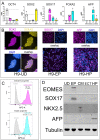
Human pluripotent stem cells efficiently form endoderm and hepatic progenitor cells in vitro. (A) RT-qPCR analysis of RNA extracted from UD, EP, HP, and ectoderm (ECT) cells displayed as delta-delta Ct values relative to HMBS “housekeeping gene product” (n = 3 biological replicates shown as individual points, bars show mean, and error bars show standard deviation). (B) Representative images of indirect immunofluorescence of UD H9 ESCs showing SOX2 (magenta), SSEA3 (yellow), DAPI (blue), and each channel overlaid; of EP cells showing SOX17 (magenta), DAPI (blue), and the two channels overlaid; and HP cells with AFP (magenta), DAPI (blue), and each channel overlaid. Scale bar denotes 100 µm. (C) Flow cytometry analysis of EP cells (top) stained with either isotype control antibody: APC conjugate (gray histogram) or anti-CXCR4: APC conjugate (magenta histogram), and HP cells (bottom) stained with either isotype control antibody: APC conjugate (gray histogram) or anti-CD99: APC conjugate (cyan histogram). Histograms show a result representative from four biological replicates; mean values for percent positive are shown ±standard deviation. (D) Western blot analysis of total protein extracted from UD, EP, CM, ECT, or HP cells and probed with antibodies directed against EOMES, SOX17, NKX2.5, AFP, and Tubulin; results shown are representative of at least two biological replicates. CM: cardiac mesoderm; DAPI: 4’,6-diamidino-2-phenylindole; EP: endoderm progenitor; HP: hepatic progenitor; UD: undifferentiated hESCs.
Figure 2.
Figure 2.
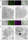
Human EP and HP cells readily endocytose SPM particles in vitro. (A) Representative result from live imaging of human EP cells with phase contrast (gray) and fluorescent imaging of nuclear-localized hrGFP (green), SPM-flash-red conjugate, and resulting overlaid images. Scale bar denotes 50 µm. (B) Electron microscopy showing two independent representative images of human EP cells shown in (A). Dark-staining objects are SPM particles; scale bar denotes 4 µm. (C) Representative result from live imaging of human HP cells with phase contrast (gray) and fluorescent imaging of nuclear-localized hrGFP (green), SPM-flash-red conjugate, and resulting overlaid images. Scale bar denotes 50 µm. (D) Electron microscopy showing two independent representative images of human HP. Dark-staining objects are SPM particles (indicated by black arrow in the image on right, while white arrowheads indicate membrane); scale bar denotes 4 µm (left) or 200 nm (right). EP: endoderm progenitor; HP: hepatic progenitor.
Figure 3.
Figure 3.
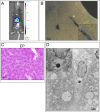
SPM-labeled human EP cells are readily detectable in mouse liver 3 h post-transplant. (A) In vivo imaging of luciferase-expressing EP cells 3 h post-transplant. (B) Fluorescent microscopy analysis of mouse liver section, from mouse shown in (A); arrowhead indicates transplanted hrGFP-expressing human EP (see inset for higher magnification); scale bar denotes 50 µm; nuclei were stained with DAPI. (C) Prussian blue staining for the presence of iron, derived from mouse liver (shown in A and B) section; scale bar denotes 100 µm. (D) Electron microscopy from mouse liver (shown in A, B, and C) with iron particle labeling transplanted human EP cell; scale bars denote 2 µm. DAPI: 4’,6-diamidino-2-phenylindole; EP: endoderm progenitor.
Figure 4.
Figure 4.
EMT is intrinsically activated in human EP and HP cells differentiated in vitro. (A) RT-qPCR analysis of RNA extracted from UD, EP, HP, and ECT cells displayed as delta-delta Ct values relative to HMBS “housekeeping gene product,” except in the case of N-Cadherin/E-Cadherin (NCAD/ECAD) where NCAD is shown relative to ECAD to determine the ratio of transcript abundances (n = 3 biological replicates shown as individual points, bars show mean, error bars show standard deviation). (B) Representative result of indirect immunofluorescence analyzed by fluorescent microscopy of human UD, EP, and HP cells stained with antibodies directed against E-Cadherin (magenta) or N-Cadherin (yellow), or stained with DAPI (blue), and each channel overlaid; scale bars denote 50 µm. Graph on the right shows fluorescent intensity ratios of N-Cadherin/E-Cadherin calculated per cell, with each point plotted representing a single cell (ns = not significant; ****P < 0.001 by Mann–Whitney and one-way ANOVA tests). ANOVA: analysis of variance; DAPI: 4’,6-diamidino-2-phenylindole; EMT: epithelial-to-mesenchymal transition; EP: endoderm progenitor; HP: hepatic progenitor; UD: undifferentiated hESCs.
Figure 5.
Figure 5.
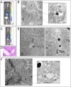
SPM-labeled human EP and HP cells are detectable in mouse liver 24 h post-transplant and exhibit features of EMT. (A) In vivo imaging of luciferase-expressing EP cells 24 h post-transplant. (B) Electron microscopy from mouse liver (shown in A) with iron particle-labeled transplanted human EP cells; scale bar denotes 1 µm on left image, with boxed region shown at higher magnification on right (scale bar denotes 500 nm). (C) In vivo imaging of luciferase-expressing HP cells 24 h post-transplant. (D) Prussian blue staining for the presence of iron, derived from mouse liver (shown in C) section; scale bar denotes 100 µm. (E) Electron microscopy from two independent lobes of mouse liver (shown in C and D) with iron particle-labeled transplanted human HP cells; scale bars denote 2 µm. (F) Electron microscopy from mouse liver (shown in C, D, and E) with iron particle-labeled transplanted human HP cells immunolabeled with Vimentin antibody and gold nanoparticle conjugate; scale bar denotes 2 µm in image on left, with boxed region shown at higher magnification on right (scale bar denotes 500 nm); arrowheads indicate gold particles. EMT: epithelial-to-mesenchymal transition; EP: endoderm progenitor; HP: hepatic progenitor.

Similar articles

Cited by

References

    1. Jorns C, Ellis EC, Nowak G, Fischler B, Nemeth A, Strom SC, Ericzon BG. Hepatocyte transplantation for inherited metabolic diseases of the liver. J Intern Med. 2012;272(3):201–223. - PubMed
    1. Strom SC, Bruzzone P, Cai H, Ellis E, Lehmann T, Mitamura K, Miki T. Hepatocyte transplantation: clinical experience and potential for future use. Cell Transplant. 2006;15(Suppl 1):S105–S110. - PubMed
    1. Horslen SP, McCowan TC, Goertzen TC, Warkentin PI, Cai HB, Strom SC, Fox IJ. Isolated hepatocyte transplantation in an infant with a severe urea cycle disorder. Pediatrics. 2003;111(6 Pt 1):1262–1267. - PubMed
    1. Gupta S, Rajvanshi P, Sokhi R, Slehria S, Yam A, Kerr A, Novikoff PM. Entry and integration of transplanted hepatocytes in rat liver plates occur by disruption of hepatic sinusoidal endothelium. Hepatology. 1999;29(2):509–519. - PubMed
    1. Baccarani U, Adani GL, Sanna A, Avellini C, Sainz-Barriga M, Lorenzin D, Montanaro D, Gasparini D, Risaliti A, Donini A, Bresadola F. Portal vein thrombosis after intraportal hepatocytes transplantation in a liver transplant recipient. Transpl Int. 2005;18(6):750–754. - PubMed

Publication types