Review
. 2021 Mar 3;28(1):15.
doi: 10.1186/s12929-021-00714-w.

Rheumatoid cachexia: the underappreciated role of myoblast, macrophage and fibroblast interplay in the skeletal muscle niche

Affiliations
Review

Rheumatoid cachexia: the underappreciated role of myoblast, macrophage and fibroblast interplay in the skeletal muscle niche

T Ollewagen et al. J Biomed Sci. .

Abstract

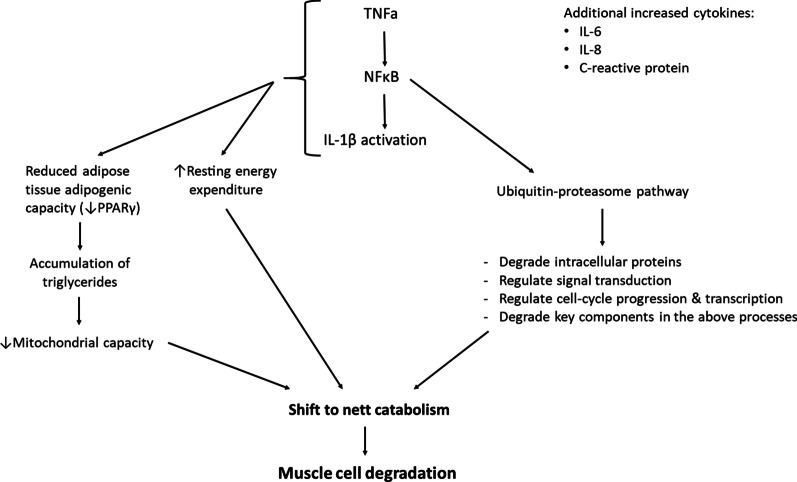
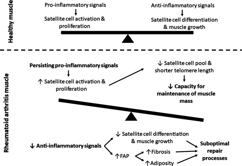
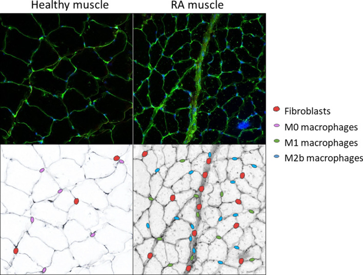
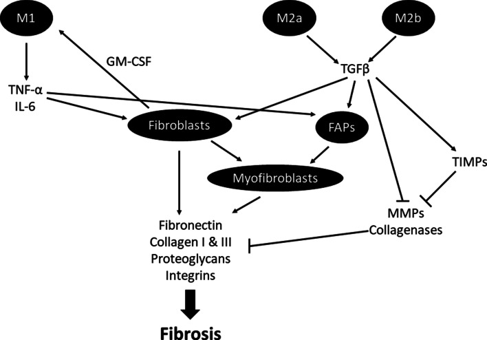
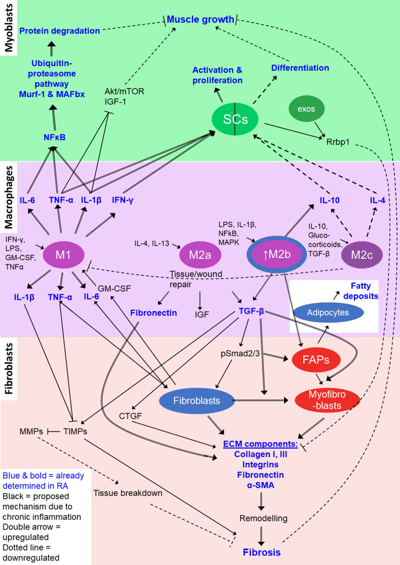
Although rheumatoid arthritis affects 1% of the global population, the role of rheumatoid cachexia, which occurs in up to a third of patients, is relatively neglected as research focus, despite its significant contribution to decreased quality of life in patients. A better understanding of the cellular and molecular processes involved in rheumatoid cachexia, as well as its potential treatment, is dependent on elucidation of the intricate interactions of the cells involved, such as myoblasts, fibroblasts and macrophages. Persistent RA-associated inflammation results in a relative depletion of the capacity for regeneration and repair in the satellite cell niche. The repair that does proceed is suboptimal due to dysregulated communication from the other cellular role players in this multi-cellular environment. This includes the incomplete switch in macrophage phenotype resulting in a lingering pro-inflammatory state within the tissues, as well as fibroblast-associated dysregulation of the dynamic control of the extracellular matrix. Additional to this endogenous dysregulation, some treatment strategies for RA may exacerbate muscle wasting and no multi-cell investigation has been done in this context. This review summarizes the most recent literature characterising clinical RA cachexia and links these features to the roles of and complex communication between multiple cellular contributors in the muscle niche, highlighting the importance of a targeted approach to therapeutic intervention.

Keywords: Arthritis; Co-culture; Extracellular matrix; Intracellular communication; M2b; Macrophage.


Conflict of interest statement

The authors confirm no conflict of interest.

Figures

Fig. 1
Fig. 1
Pathways of proinflammatory cytokine-induced protein degradation in rheumatoid cachexia. (ref [, –45]). TNFα tumor necrosis factor-α, NFκB  nuclear factor kappa-light chain enhancer of activated B cells, IL-1β  interleukin-1β, PPARγ  peroxisome proliferator activated receptor-gamma
Fig. 2
Fig. 2
The imbalance between pro- and anti-inflammatory signalling in RA rodent skeletal muscle, and the resulting influence on muscle repair and growth. FAP  fibro-adipogenic progenitor cell
Fig. 3
Fig. 3
Representative images suggesting the presence of different cell types in healthy versus rheumatoid arthritis skeletal muscle. Fluorescent images from a RA rodent model (study execution described in Oyenihi et al., 2019 [38]; staining method described in Additional file 1) indicate clear cachexia and increased fibrosis between muscle fibres. Black and white images indicative of the authors prediction of greater presence of macrophages (M1 and M2b) and fibroblasts in RA
Fig. 4
Fig. 4
Summary of the interaction between macrophages, fibroblasts and FAPs in the development of tissue fibrosis. TNF-α  tumor necrosis factor-alpha, IL-6  interleukin-6, TGFβ  transforming growth factor-beta, FAPs  fibro-adipogenic progenitor cells, TIMPs  tissue inhibitor of metalloproteinase, MMPs  metalloproteinases
Fig. 5
Fig. 5
Summary of the cellular interactions in chronic inflammation leading to a decline in muscle growth. The incomplete switch from M1 to M2c results in a greater presence of M2b macrophages which present both impaired pro- and impaired anti-inflammatory properties, often resulting in enhanced deposition of ECM components, and impaired satellite cell function. This also results in a reduced presence of M2c macrophages. The mechanisms and pathways presented are based on chronic inflammatory microenvironments (proposed in RA), with blue indicating mechanisms confirmed in studies of RA. Different colours indicate different cell focus areas; green = muscle niche; purple = inflammatory system; red = fibroblasts and fibrosis. Increased signalling are indicated by double-line arrows, while dotted line arrows indicate decreased signalling. SCs  satellite cells, IGF  insulin-like growth factor, Murf-1  muscle ring finger protein-1, Mafbx  muscle atrophy f-box, exos  exosomes, Rrbp1  ribosome binding protein-1, IL  interleukin, TNF-α  tumor necrosis factor-α, IFN-γ interferon-γ, LPS  lipopolysaccharides, GM-CSF  granulocyte-monocyte colony-stimulating factor, TGF-β  transforming growth factor-β, TIMP  tissue inhibitor of metalloproteinase, MMP  matrix metalloproteinase, CTGF  connective tissue growth factor, FAPs  fibro-adipogenic progenitor cells, ECM  extracellular matrix, α-SMA  α-smooth muscle actin

References

    1. Gaffo A, Saag KG, Curtis JR. Treatment of rheumatoid arthritis. Clin Rev. 2006;63:2451–2465. - PubMed
    1. Majithia V, Geraci SA. Rheumatoid arthritis: diagnosis and management. Am J Med. 2007;120:936–939. doi: 10.1016/j.amjmed.2007.04.005. - DOI - PubMed
    1. Lemmey AB, Wilkinson TJ, Clayton RJ, Sheikh F, Whale J, Jones HSJ, et al. Tight control of disease activity fails to improve body composition or physical function in rheumatoid arthritis patients. Rheumatol (United Kingdom) 2016;55(10):1736–1745. - PubMed
    1. Filippin LI, Teixeira VN, Viacava PR, Lora PS, Xavier LL, Xavier RM. Temporal development of muscle atrophy in murine model of arthritis is related to disease severity. J Cachexia Sarcopenia Muscle. 2013;4(3):231–238. doi: 10.1007/s13539-013-0102-1. - DOI - PMC - PubMed
    1. Andonian BJ, Huffman KM. Skeletal muscle disease in rheumatoid arthritis: the center of cardiometabolic comorbidities? Curr Opin Rheumatol. 2020;32(3):297–306. doi: 10.1097/BOR.0000000000000697. - DOI - PubMed