. 2021 Mar 3;7(10):eabc7606.
doi: 10.1126/sciadv.abc7606. Print 2021 Mar.

Durable memories and efficient neural coding through mnemonic training using the method of loci

Affiliations

Durable memories and efficient neural coding through mnemonic training using the method of loci

I C Wagner et al. Sci Adv. .

Abstract

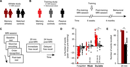
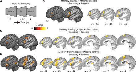
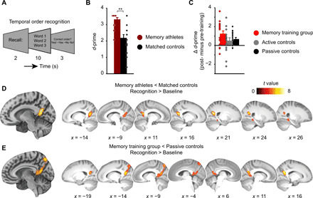
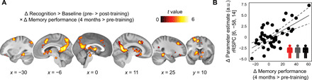
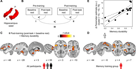
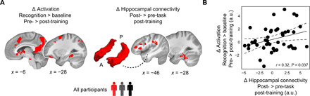
Mnemonic techniques, such as the method of loci, can powerfully boost memory. We compared memory athletes ranked among the world's top 50 in memory sports to mnemonics-naïve controls. In a second study, participants completed a 6-week memory training, working memory training, or no intervention. Behaviorally, memory training enhanced durable, longer-lasting memories. Functional magnetic resonance imaging during encoding and recognition revealed task-based activation decreases in lateral prefrontal, as well as in parahippocampal and retrosplenial cortices in both memory athletes and participants after memory training, partly associated with better performance after 4 months. This was complemented by hippocampal-neocortical coupling during consolidation, which was stronger the more durable memories participants formed. Our findings advance knowledge on how mnemonic training boosts durable memory formation through decreased task-based activation and increased consolidation thereafter. This is in line with conceptual accounts of neural efficiency and highlights a complex interplay of neural processes critical for extraordinary memory.


Figures

Fig. 1
Fig. 1. Study design, procedures, and results from the free recall tests.
(A) In the athlete study, we tested memory athletes (n = 17) and compared them to matched controls (n = 16) during a single MRI session. (B) Participants of the training study were pseudo-randomized into three groups after an initial MRI session (pre-training): the memory training group (n = 17), active controls (n = 16), and passive controls (n = 17). Participants returned to the laboratory for a second MRI session (post-training) and took part in a behavioral retest after 4 months. (C) General structure of MRI sessions: baseline resting-state period (8 min), word list encoding and temporal order recognition tasks (10 min each), post-task resting-state period (8 min), immediate free recall test (5 + 5 min and 20 min post-MRI), and delayed free recall test after 24 hours (5 + 5 min; only completed by participants of the training study, dashed frame). (D) Training study: Change in the number of forgotten/weak/durable words from pre- to post-training sessions. Note that only weak and durable memories were included in the analysis (marked in bold). **P < 0.0001. (E) Athlete study: Free recall performance (20 min). **P = 0.0005. (D and E) Error bars reflect the SEM. See also Table 2 for an overview of free recall performance across the groups.
Fig. 2
Fig. 2. Activation changes during word list encoding.
(A) Word list encoding task: Participants studied previously unstudied words during each MRI session. (B) Athlete study: Brain activation during encoding (encoding > baseline) is decreased in memory athletes compared to matched controls. (C and D) Training study: Brain activation is significantly decreased in the memory training group after training when compared to (C) active or (D) passive controls (group × session interactions; see table S1 for main effects and table S2 for a comparison between active and passive controls). Results are shown at P < 0.05 family-wise error (FWE)–corrected at cluster level (cluster-defining threshold P < 0.001). LH, left hemisphere.
Fig. 3
Fig. 3. D-prime and activation changes during temporal order recognition.
(A) After word list encoding, word triplets were presented in the same or a different order as studied previously and participants were asked to judge the order. (B) Athlete study: Recognition performance (d-prime) for memory athletes and matched controls. **P < 0.001. (C) Training study: Change in d-prime (from pre- to post-training sessions) across the groups (main effect of group, P = 0.133). Error bars (B and C) reflect the SEM. See also Table 2 for an overview of recognition performance across the groups. (D) Athlete study: Brain activation during temporal order recognition (recognition > baseline) is decreased in memory athletes compared to matched controls (see Results for MNI coordinates). (E) Training study: Brain activation is significantly decreased in the memory training group after training when compared to passive controls (group × session interaction; see table S3). Results are shown at P < 0.05 FWE-corrected at cluster level (cluster-defining threshold P < 0.001).
Fig. 4
Fig. 4. Activation changes during temporal order recognition and association with memory performance at the 4-month retest.
(A) Training study: Decreases in brain activation (recognition > baseline) from before to after training (pre- > post-training) that positively scaled with the change in free recall performance (referred to “memory performance” in the figure) from the pre-training session (20 min post-MRI) to the retest after 4 months (covariate of interest). Results are shown at P < 0.05 FWE-corrected at cluster level (cluster-defining threshold P < 0.001; see also table S4). (B) The scatterplot shows the relationship between the change in parameter estimates [arbitrary units (a.u.)] from the pre- to post-training sessions, extracted from the global maximum (right retrosplenial cortex, rRSPC; 8-mm sphere around MNI peak coordinate, x = 6, y = −58, z = 14), and the change in memory performance (4-month retest minus pre-training20 min). Given the clear inferential circularity, we would like to highlight that this plot serves visualization purposes only, solely illustrating the direction of association between the brain-behavior relationship.
Fig. 5
Fig. 5. Hippocampal connectivity at rest and association with memory durability.
(A) Bilateral anatomical hippocampus seed used for whole-brain connectivity analysis. A, anterior, P, posterior. (B) Training study: Schematic of the analysis steps performed. We tested hippocampal connectivity increases (post-task > baseline rest) during the pre- (I) and post-training sessions (II) and investigated the increase in consolidation-related coupling from pre- to post-training sessions (III; [post-task > baseline rest]post > [post-task > baseline rest]pre). Analysis of data from the athlete study involved a single MRI session (post-task > baseline), which is not depicted here. (C) Training study: Hippocampal-neocortical connectivity increases from baseline to post-task rest during the post-training session positively scaled with the proportion of durable memories formed (i.e., memory durability) across all participants (see also table S5). (D) Follow-up analyses revealed that these effects were specifically driven by connectivity changes in the memory training group but were not present in passive or active controls (see results S9 and table S6). Given the clear inferential circularity, we would like to highlight that the scatterplot (E) serves visualization purposes only, solely illustrating the direction of association between the brain-behavior relationship. All results are shown at P < 0.05 FWE-corrected at cluster level (cluster-defining threshold P < 0.001).
Fig. 6
Fig. 6. Relation between task-based activation decreases and hippocampal connectivity at rest.
(A) (Left) We created a whole-brain binary mask centered on the significant activation effects obtained during temporal order recognition (Fig. 4; based on a sample of n = 45) and extracted the raw change in activation per participant (i.e., using the contrast pre- minus post-training, recognition > baseline). (Right) We created a whole-brain binary mask centered on the significant connectivity effects obtained during post-task resting state after training (Fig. 5; based on a sample of n = 49) and extracted the raw change in hippocampal connectivity per participant (i.e., using the contrast post- minus pre-task resting state, during the post-training session). The bilateral hippocampal seed is schematically indicated (A, anterior; P, posterior). (B) Correlational analysis across all participants of the training study. Larger activation decreases (i.e., more positive values pre-training) were coupled to larger increases in hippocampal connectivity after training.

References

    1. F. Yates, The Art of Memory (Routledge & Kegan, 1966).
    1. J. Foer, Moonwalking with Einstein (Penguin Books, 2011).
    1. Dresler M., Shirer W. R., Konrad B. N., Müller N. C. J., Wagner I. C., Fernández G., Czisch M., Greicius M. D., Mnemonic training reshapes brain networks to support superior memory. Neuron 93, 1227–1235.e6 (2017). - PMC - PubMed
    1. Maguire E. A., Valentine E. R., Wilding J. M., Kapur N., Routes to remembering: The brains behind superior memory. Nat. Neurosci. 6, 90–95 (2003). - PubMed
    1. Fellner M.-C., Volberg G., Wimber M., Goldhacker M., Greenlee M. W., Hanslmayr S., Spatial Mnemonic encoding: Theta power decreases and medial temporal lobe BOLD increases co-occur during the usage of the method of loci. eNeuro 3, ENEURO.0184-16.2016 (2016). - PMC - PubMed

Publication types

LinkOut - more resources