

Mantis shrimp-inspired organic photodetector for simultaneous hyperspectral and polarimetric imaging
- PMID: 33658196
- PMCID: PMC7929508
- DOI: 10.1126/sciadv.abe3196
Mantis shrimp-inspired organic photodetector for simultaneous hyperspectral and polarimetric imaging
Abstract
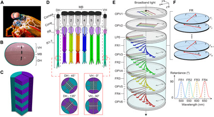
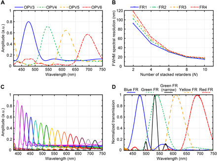
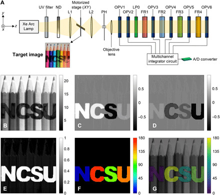
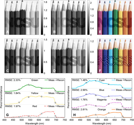
Combining hyperspectral and polarimetric imaging provides a powerful sensing modality with broad applications from astronomy to biology. Existing methods rely on temporal data acquisition or snapshot imaging of spatially separated detectors. These approaches incur fundamental artifacts that degrade imaging performance. To overcome these limitations, we present a stomatopod-inspired sensor capable of snapshot hyperspectral and polarization sensing in a single pixel. The design consists of stacking polarization-sensitive organic photovoltaics (P-OPVs) and polymer retarders. Multiple spectral and polarization channels are obtained by exploiting the P-OPVs' anisotropic response and the retarders' dispersion. We show that the design can sense 15 spectral channels over a 350-nanometer bandwidth. A detector is also experimentally demonstrated, which simultaneously registers four spectral channels and three polarization channels. The sensor showcases the myriad degrees of freedom offered by organic semiconductors that are not available in inorganics and heralds a fundamentally unexplored route for simultaneous spectral and polarimetric imaging.
Copyright © 2021 The Authors, some rights reserved; exclusive licensee American Association for the Advancement of Science. No claim to original U.S. Government Works. Distributed under a Creative Commons Attribution NonCommercial License 4.0 (CC BY-NC).
Figures
References
-
- Tyo J. S., Goldstein D. L., Chenault D. B., Shaw J. A., Review of passive imaging polarimetry for remote sensing applications. Appl. Optics 45, 5453–5469 (2006). - PubMed
-
- Y. Zhao, C. Yi, S. G. Kong, Q. Pan, Y. Cheng, Multi-Band Polarization Imaging and Applications (Advances in Computer Vision and Pattern Recognition, Springer Berlin Heidelberg, 2016).
-
- Pu Y., Wang W. B., Tang G. C., Zeng F., Achilefu S., Vitenson J. H., Sawczuk I., Peters S., Lombardo J. M., Alfano R. R., Spectral polarization imaging of human prostate cancer tissue using a near-infrared receptor-targeted contrast agent. Technol. Cancer Res. Treat. 4, 429–436 (2005). - PubMed
-
- Oka K., Kato T., Spectroscopic polarimetry with a channeled spectrum. Opt. Lett. 24, 1475–1477 (1999). - PubMed
LinkOut - more resources
Full Text Sources
Other Literature Sources
