. 2021 Mar 3;7(10):eabf5814.
doi: 10.1126/sciadv.abf5814. Print 2021 Mar.

Cryo-EM structures of excitatory amino acid transporter 3 visualize coupled substrate, sodium, and proton binding and transport

Affiliations

Cryo-EM structures of excitatory amino acid transporter 3 visualize coupled substrate, sodium, and proton binding and transport

Biao Qiu et al. Sci Adv. .

Abstract

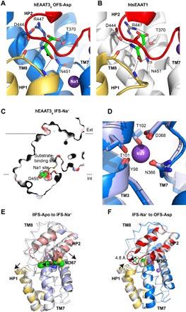
Human excitatory amino acid transporter 3 (hEAAT3) mediates glutamate uptake in neurons, intestine, and kidney. Here, we report cryo-EM structures of hEAAT3 in several functional states where the transporter is empty, bound to coupled sodium ions only, or fully loaded with three sodium ions, a proton, and the substrate aspartate. The structures suggest that hEAAT3 operates by an elevator mechanism involving three functionally independent subunits. When the substrate-binding site is near the cytoplasm, it has a remarkably low affinity for the substrate, perhaps facilitating its release and allowing the rapid transport turnover. The mechanism of the coupled uptake of the sodium ions and the substrate is conserved across evolutionarily distant families and is augmented by coupling to protons in EAATs. The structures further suggest a mechanism by which a conserved glutamate residue mediates proton symport.


Figures

Fig. 1
Fig. 1. Alternating access mechanism of hEAAT3g.
(A) hEAAT3g (open squares) and wild-type hEAAT3 (solid squares) show similar l-asp uptake in oocytes. Measured currents are plotted as fractions of the maximal observed current. (B) Transient currents mediated by hEAAT3g reconstituted into solid-supported membranes. Measured peak currents are plotted as fractions of the maximal observed peak current. Currents were measured in buffers containing nonpermeant phosphate (open circles) and permeant chloride (solid squares) anions. (C) Observed populations of hEAAT3g trimers with different configurations of protomers in the OFS (O) and IFS (I), as indicated below the graph (black columns), are consistent with binomial distributions with OFS probability of 19% (gray columns). The analysis is based on the cryo-EM imaging data for hEAAT3g in the presence of 200 mM NaCl and 1 mM l-asp. (D) Structures of single hEAAT3g protomers in the IFS-Na+ (left) and OFS-Asp (right) states. The scaffold domains are pink and beige, and the transport domains are in shades of blue. Structurally symmetric HP1 and HP2 are yellow and red, respectively. Substrate and ions are shown as spheres and colored by atom type. Below are the close-up views of the substrate-binding sites. The gray mesh objects are density maps contoured at 5.5 σ for L-asp (left) and 4.5 σ for the protein (right). (E) Interactions between HP2 (red spheres) and scaffold domain (beige surface) for the inward-facing hEAAT3g bound to Na+ ions (left), ASCT2 bound to Na+ ions [Protein Data Bank (PDB) accession code 6rvx, middle], and ASCT2-Gln bound to glutamine (PDB accession code 6gct, right). Dotted blue ellipses highlight the locations of the substrate-binding site for reference. The rest of the transport domains are shown as blue ribbons. The close-up views are below the panels.
Fig. 2
Fig. 2. Substrate and sodium coupling in hEAAT3g.
l-Asp binds differently to hEAAT3g (A) and thermally stabilized human EAAT1 (htsEAAT1, PDB accession code 5 lm4) (B). While the electron density for l-asp is a little ambiguous in htsEAAT1 structure, the substrate position and coordination is identical to GltPh and GltTk, lending confidence to the model. Interactions between l-asp and EAATs are shown as dashed lines. HP1 and HP2 are colored yellow and red, respectively; l-asp is colored green; and TM7 and TM8 are colored blue (hEAAT3g) and white (htsEAAT1), respectively. (C) Na1 is solvent accessible. hEAAT3g is shown as a thin slice taken through the Na1 site. The Na+-coordinating residue D455 is shown as spheres and colored by atom type. (D) Close-up view of the Na3 site in hEAAT3g OFS-Asp (marine) and IFS-Na+ (light blue). (E and F) Conformational changes within the transport domains transitioning from the IFS-Apo to IFS-Na+ state (E) and IFS-Na+ to OFS-Asp state (F). The transport domains are white and shades of blue for the starting and ending states, respectively, with HP1 in yellow and HP2 in salmon. M367 is shown as spheres and colored by atom type in all states. The arrows indicate conformational changes from IFS-Apo to IFS-Na+ (E) and IFS-Na+ to OFS-Asp (F).
Fig. 3
Fig. 3. The proton coupling mechanism of hEAAT3g.
Close-up views of R447 and E374 in the OFS-Asp state (A), IFS-Na+ state (B), and IFS-Apo state (C). Key interactions are shown as red dashed lines; density of residues is shown as mesh; the contour levels of the maps are below the panels. HP1 and HP2 are yellow and red, respectively. TM7 and TM8 are marine, light blue, and white in (A), (B), and (C), respectively. l-Asp is green. Position of R447 and E374 relative to the membrane and their solvent accessibility in the OFS-Asp state (D), IFS-Na+ (E), and IFS-Apo state (F). hEAAT3g is shown as a surface sliced approximately through the substrate-binding site and viewed in the membrane plane. Dashed ellipses mark the scaffold domains. R447 and E374 are shown as spheres.

References

    1. Vandenberg R. J., Ryan R. M., Mechanisms of glutamate transport. Physiol. Rev. 93, 1621–1657 (2013). - PubMed
    1. Grewer C., Gameiro A., Rauen T., SLC1 glutamate transporters. Pflugers Arch. 466, 3–24 (2014). - PMC - PubMed
    1. Bjorn-Yoshimoto W. E., Underhill S. M., The importance of the excitatory amino acid transporter 3 (EAAT3). Neurochem. Int. 98, 4–18 (2016). - PMC - PubMed
    1. Kanai Y., Hediger M. A., Primary structure and functional characterization of a high-affinity glutamate transporter. Nature 360, 467–471 (1992). - PubMed
    1. Rothstein J. D., Martin L., Levey A. I., Dykes-Hoberg M., Jin L., Wu D., Nash N., Kuncl R. W., Localization of neuronal and glial glutamate transporters. Neuron 13, 713–725 (1994). - PubMed

Publication types

LinkOut - more resources