Review
. 2021 Apr 9;8(2):ENEURO.0337-20.2021.
doi: 10.1523/ENEURO.0337-20.2021. Print 2021 Mar-Apr.

The Kainic Acid Models of Temporal Lobe Epilepsy

Affiliations
Review

The Kainic Acid Models of Temporal Lobe Epilepsy

Evgeniia Rusina et al. eNeuro. .

Abstract

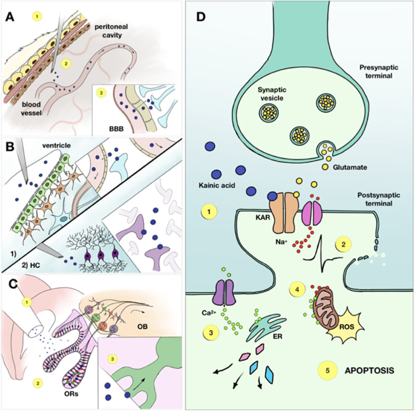
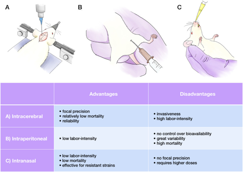
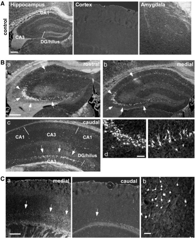
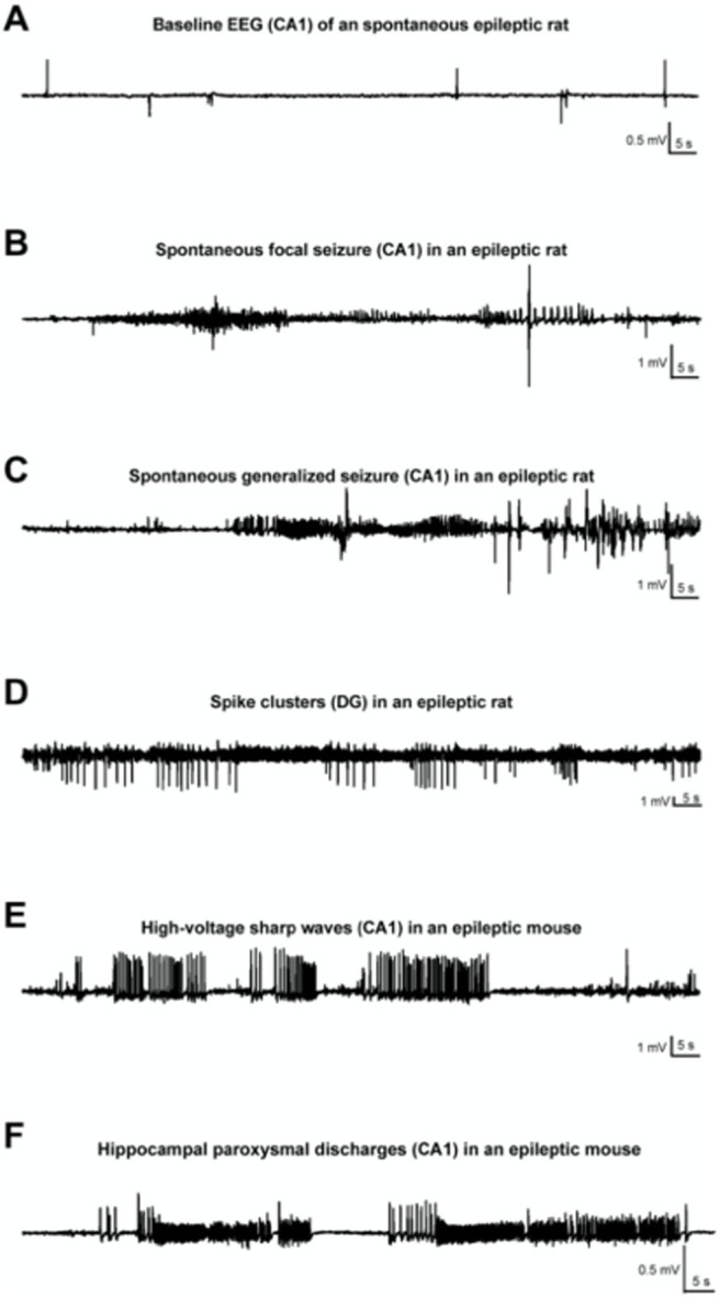
Experimental models of epilepsy are useful to identify potential mechanisms of epileptogenesis, seizure genesis, comorbidities, and treatment efficacy. The kainic acid (KA) model is one of the most commonly used. Several modes of administration of KA exist, each producing different effects in a strain-, species-, gender-, and age-dependent manner. In this review, we discuss the advantages and limitations of the various forms of KA administration (systemic, intrahippocampal, and intranasal), as well as the histologic, electrophysiological, and behavioral outcomes in different strains and species. We attempt a personal perspective and discuss areas where work is needed. The diversity of KA models and their outcomes offers researchers a rich palette of phenotypes, which may be relevant to specific traits found in patients with temporal lobe epilepsy.

Keywords: EEG; hippocampus; kainic acid; mice models of temporal lobe epilepsy.


Figures

Figure 1.
Figure 1.
The complex mechanism of KA-induced neuronal damage includes a sequence of events and its outcome varies depending on the administration route. A, The intraperitoneal route of KA administration is realized through an injection of the drug into the peritoneal cavity (1). The molecules then get absorbed by blood vessels (2) and travel to the brain, where they pass the blood-brain barrier via passive diffusion (3). B, The intracerebral administration can be performed in various zones but the most common are intraventricular (1) and intrahippocampal (2). In case of intraventricular injection, the drug molecules diffuse directly into the cells surrounding the ventricle wall and another part is taken by the blood vessels to be distributed throughout the entire brain. Intrahippocampal administration, on the contrary, provides a more localised damage, as KA molecules activate KA receptors in the hippocampus at the site of injection. C, The intranasal route starts with the injection of KA in the nasal cavity (1), where the molecules are absorbed by receptors of the olfactory epithelium (2), from where they travel through the olfactory pathway to the hippocampus and other areas of the brain (3). D, KA, once having reached the brain tissue, initiates a cascade of events First, it binds to the KARs, causing membrane depolarization and cell firing (1). Excessive firing can lead to osmotic imbalance and, eventually, rupture of the postsynaptic membrane (2). At the same time, influx of calcium into the cell results in multiple enzymes activation, such as phospholipase, endonucleases and proteases, all of which damage various cell structures (3). Additional effect of an increased intracellular Ca2+ concentration is mitochondrial disfunction, and excessive production of reactive oxygen species (4). All these mechanisms potentiate each other and terminate in apoptosis (5). BBB — blood-brain barrier, ER — endoplasmatic reticulum, HC — hippocampus, KAR — kainic acid receptor, OB — olfactory bulb, ORs — olfactory receptors, ROS — reactive oxygen species.
Figure 2.
Figure 2.
Advantages and disadvantages of KA administration routes. Various points should be taken into consideration, including age, sex and strain of an animal. A, The main advantage of the intracerebral administration route is focal precision; this method is widely used, despite of its invasiveness and labor-intensity. B, The intraperitoneal injection is easy to perform, but might result in high mortality, along with large outcome variability and uncontrolled tissue uptake. C, The intranasal route leads to low mortality rates and works for resistant strains, but lacks focal precision.
Figure 3.
Figure 3.
Neuropathological alterations in hippocampal and cortical areas followed by intra-amygdaloid KA administration, FluoroJade B (FjB) staining. A, Hippocampus, cortex and amygdala of a control mouse. Absence of FjB-positive cells. B, Images of ipsilateral hippocampus of a KA-treated mouse at anterior, median and posterior levels. FjB-positive cells are indicated by arrows. C, Representative images of ipsilateral temporal cortex of a KA-treated mouse. (Mouri et al., 2008).
Figure 4.
Figure 4.
Various patterns of EEG-activity during KA-induced chronic epilepsy. A, Baseline recording from CA1 of an epileptic rat. Note the occurrence of interictal spikes. B, Recording of a spontaneous focal seizure in CA1. C. A secondary generalized convulsive seizure in an epileptic rat. D, Spike clusters originating from the dentate gyrus 7 weeks post-SE. E, High-voltage sharp waves in the epileptic focus (CA1) of a mouse, several weeks post-SE. F, Hippocampal paroxysmal discharges (HPDs) in CA1 of an epileptic mouse. (Klee et al., 2017).
Figure 5.
Figure 5.
Seizure progression over time. A, Increase in seizure frequency over 14 weeks post-injection. The latent period is reflected in stage 1, stage 2 represents the “slow growth phase”, stage 3 is characterised by an exponential growth until reaching the steady stage 4. B, Actual seizure frequency plotted on the graph. Data obtained from the 9 animals. C, Normalised seizure frequency, same cohort. (Williams et al., 2009).

References

    1. Aicardi J, Chevrie JJ (1970) Convulsive status epilepticus in infants and children. Epilepsia 11:187–197. 10.1111/j.1528-1157.1970.tb03880.x - DOI - PubMed
    1. Andermann F, Kobayashi E, Andermann E (2005) Genetic focal epilepsies: state of the art and paths to the future. Epilepsia 46:61–67. 10.1111/j.1528-1167.2005.00361.x - DOI - PubMed
    1. Anthony Altar C, Baudry M (1990) Systemic injection of kainic acid: gliosis in olfactory and limbic brain regions quantified with [3H]PK 11195 binding autoradiography. Exp Neurol 109:333–341. 10.1016/S0014-4886(05)80024-X - DOI - PubMed
    1. Araki T, Simon RP, Taki W, Lan J-Q, Henshall DC (2002) Characterization of neuronal death induced by focally evoked limbic seizures in the C57BL/6 mouse. J Neurosci Res 69:614–621. 10.1002/jnr.10356 - DOI - PubMed
    1. Arabadzisz D, Antal K, Parpan F, Emri Z, Fritschy JM (2005) Epileptogenesis and chronic seizures in a mouse model of temporal lobe epilepsy are associated with distinct EEG patterns and selective neurochemical alterations in the contralateral hippocampus. ExpNeurol 194:76–90. - PubMed