. 2021 Apr 26;95(10):e00008-21.
doi: 10.1128/JVI.00008-21. Epub 2021 Mar 3.

Interaction of NEP with G Protein Pathway Suppressor 2 Facilitates Influenza A Virus Replication by Weakening the Inhibition of GPS2 to RNA Synthesis and Ribonucleoprotein Assembly

Affiliations

Interaction of NEP with G Protein Pathway Suppressor 2 Facilitates Influenza A Virus Replication by Weakening the Inhibition of GPS2 to RNA Synthesis and Ribonucleoprotein Assembly

Wenxiao Gong et al. J Virol. .

Abstract

The nuclear export protein (NEP) serves multiple functions in the life cycle of influenza A virus (IAV). Identifying novel host proteins that interact with NEP and understanding their functions in IAV replication are of great interest. In this study, we screened and confirmed the direct interaction of G protein pathway suppressor 2 (GPS2) with NEP through a yeast two-hybrid screening assay and glutathione S-transferase-pulldown and co-immunoprecipitation assays. Knockdown or knockout of GPS2 enhanced IAV titers, whereas overexpression of GPS2 impaired IAV replication, demonstrating that GPS2 acted as a negative host factor in IAV replication. Meanwhile, GPS2 inhibited viral RNA synthesis by reducing the assembly of IAV polymerase. Interestingly, IAV NEP interacted with GPS2 and mediated its nuclear export, thereby activated the degradation of GPS2. Thus, NEP-GPS2 interaction weakened the inhibition of GPS2 to viral polymerase activity and benefited virus replication. Overall, this study identified the novel NEP-binding host partner GPS2 as a critical host factor to participate in IAV replication. These findings provided novel insights into the interactions between IAV and host cells, revealing a new function for GPS2 during IAV replication.Importance: NEP is proposed to play multiple biologically important roles in the life cycle of IAV, which largely relies on host factors by interaction. Our study demonstrated that GPS2 could reduce the interaction between PB1 and PB2 and interfere with vRNP assembly. Thus, GPS2 inhibited the RNA synthesis of IAV and negatively regulated its replication. Importantly, IAV NEP interacted with GPS2 and mediated the nuclear export of GPS2, thereby activated the degradation of GPS2. Thus, NEP-GPS2 interaction weakened the inhibition of GPS2 to viral polymerase activity and benefited virus replication.


Figures

FIG 1
FIG 1
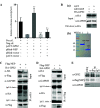
NEP interacted with GPS2. (A) NEP bound GPS2 in the MTH assay. HEK293T cells were cotransfected with pACT-GPS2, pBIND-NEP, and pG5luc, and then cell lysates were subjected to luciferase activity assays via MTH 24 h later. pBIND-ID and pACT-MyoD served as positive controls, and pACT and pBIND served as negative controls. The results are presented as the mean ± SD of three independent experiments (*, P < 0.05; **, P < 0.01; n.s., not significant; all by two-tailed Student's t test). (B) GST pulldown assay showing the direct interaction between NEP and GPS2. Lysates of HEK293T cells transfected with HA-GPS2 were incubated with an equal amount of GST or GST-NEP bound to glutathione-Sepharose 4B beads (b). Bound proteins were analyzed by Western blotting with an anti-HA monoclonal antibody (a). (C and D) Coimmunoprecipitation of HA-GPS2 and Flag-NEP in HEK293T cells. HEK293T cells were cotransfected with HA-GPS2 and Flag-NEP. Cell lysates were immunoprecipitated (IP) and immunoblotted (IB) with specific antibodies as indicated. (E) Coimmunoprecipitation of Flag-NEP in A549 cells. A549 cells were infected with WD-Flag-NEP virus at an MOI of 0.01, and cells were lysed 24 h postinfection. Cell lysates were IP and IB with specific antibodies as indicated.
FIG 2
FIG 2
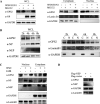
Effect of GPS2 on IAV replication. (A and B) A549 cells were transfected with sictl or siGPS2 for 24 h, followed by infection with the A/WSN/33 (WSN/H1N1) (A) or A/chicken/Hubei/327/2004 (DW/H5N1) (B) virus at an MOI of 0.01. Supernatants of the cell culture were collected at different times postinfection as indicated and assayed for virus titers by TCID50. (C and D) Effect of siGPS2 on A549 cell viability. A549 cells were transfected with siGPS2 or negative-control sictl. Cell viability was measured by cell counting kit 8 (CCK-8) assay at the indicated time points posttransfection. (E and F) Whole-cell lysates were assayed by Western blotting with the indicated antibodies. (G) GPS2-KO A549 cells (A549GPS2−/−) or empty retrovirus-transduced control A549 cells (A549KOCK) infected with the A/WSN/33 (WSN/H1N1) at an MOI of 0.01. Supernatants of the cell culture were collected at different times postinfection as indicated and assayed for virus titers by TCID50. (H) HA-GPS2 stable-expressed A549 cells (A549GPS2+) or empty retrovirus-transduced control A549 cells (A549CK) infected with A/WSN/33(WSN/H1N1) at an MOI of 0.01. Supernatants of the cell culture were collected at different times postinfection as indicated and assayed for virus titers by TCID50. For all experiments, results are presented as the mean ± SD from three independent experiments (*, P < 0.05; **, P < 0.01; n.s., not significant; all by two-tailed Student's t test).
FIG 3
FIG 3
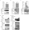
Influenza virus infection induced the degradation of GPS2 protein. (A and B) A549 cells were infected with A/WSN/33 (WSN/H1N1) or A/chicken/Hubei/327/2004 (DW/H5N1) at MOIs of 0, 0.01, 0.1, and 1, respectively. Samples were collected at 24 h postinfection. The levels of NP and GPS2 mRNA were determined by qRT-PCR. The viral RNA levels were normalized to the GAPDH level (means ± SD from three independent experiments) (*, P < 0.05; **, P < 0.01; all by two-tailed Student's t test) (A), and whole-cell lysates were assayed by Western blotting with the indicated antibodies. GAPDH was used as a loading control (B). (C) Half-lives of GPS2 in uninfected and infected A549 cells were examined. A549 cells uninfected or infected with WSN viruses (MOI = 1) were treated with CHX (100 μg/ml) at 6 h postinfection. At the indicated times after treatment, cells were harvested, and cell extracts were prepared for Western blotting to analyze GPS2 protein levels. (D) GPS2 levels shown in panel C were quantitated by band intensities analyzed using ImageJ (NIH) and normalized to GAPDH levels. (E) A549 cells infected with WSN viruses (MOI = 1) were treated with DMSO, MG132 (10 μM), or CQ (50 μM). At the indicated times, cells were harvested, and cell extracts were prepared for Western blotting using the indicated antibodies.
FIG 4
FIG 4
GPS2 degradation caused by IAV infection associated with NEP-mediated nuclear export. (A) A549 cells infected with A/WSN/33 (WSN/H1N1) at an MOI of 1 in the presence of MG132 (10 nM) and treated with DMSO as control. Cells were harvested at 24 hpi and subjected to nucleus and cytoplasmic fractions. (B) Wild-type A549 cells were infected with the A/WSN/33 (WSN/H1N1) at an MOI of 5. Samples were harvested at indicated time points postinfection and subjected to nucleus (N) and cytoplasmic (C) fractions. (C) A549 cells infected with A/WSN/33 (WSN/H1N1) at MOI of 1 in the presence of LMB (11 nM). Cells were harvested at 24 hpi and subjected to nucleus and cytoplasmic fractions. (D) A549 cells transfected with Flag-NEP or p3×Flag vector and harvested at 24 h posttransfection. Protein levels were assayed by Western blotting with the indicated antibodies. Lamin B1 was served as a nucleus loading control and marker, and GAPDH served as a cytosolic loading control and marker.
FIG 5
FIG 5
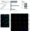
NEP mediated the nuclear export of GPS2. (A) Diagram of NEP truncated mutants, and the interaction strength with full-length GPS2 was assayed via MTH, respectively. Results are presented as the mean ± SD of three independent experiments (*, P < 0.05; **, P < 0.01; n.s., not significant; all by two-tailed Student's t test). (B) Interaction regions were confirmed by immunoprecipitation. NEP (aa 12 to 21) and NEP (aa 31 to 41) were chimeric expressed with EGFP. HEK293T cells were cotransfected with indicated plasmids, and cell lysates were immunoprecipitated and immunoblotted with specific antibodies as indicated. (C) NEP NES1 (aa 12 to 21) and NES2 (aa 31 to 41) were chimeric expressed with EGFP in pEGFP-C1 in the presence of DMSO or LMB in HeLa cells. Cells were fixed at 24 h posttransfection, and DAPI was used to stain the nucleus (blue). (D) Colocalization of GPS2 and NEP in transfected cells. HeLa cells cultured on slides were cotransfected with HA-GPS2 and/or Flag-NEP and treated with DMSO or LMB (11 nM) as indicated. HeLa cells were fixed at 24 h posttransfection and stained for HA-GPS2 (red) and NEP (green) by using the anti-HA mouse antibodies and anti-NEP rabbit antibodies, followed by immunostaining with Alexa Fluor 594-conjugated AffiniPure goat anti-mouse secondary antibodies and Alexa Fluor 488-conjugated AffiniPure goat anti-rabbit antibodies. DAPI was used to stain the nucleus (blue).
FIG 6
FIG 6
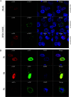
IAV infection mediated the nuclear export of GPS2. (A) HeLa cells cultured on slides were transfected with HA-GPS2 or pCAGGS-HA vector and then infected with A/WSN/33 (WSN/H1N1) at an MOI of 1 as indicated 12 h posttransfection. HeLa cells were fixed at 12 h postinfection and stained for HA-GPS2 (red) and NEP (green) by using anti-HA mouse antibodies and anti-NEP rabbit antibodies. (B) A549 cells cultured on slides were transfected with HA-GPS2 and then infected with A/WSN/33 (WSN/H1N1) at an MOI of 1 as indicated. At 3, 6, or 9 h postinfection, cells were fixed and stained for HA-GPS2 (red) and NP (green) by using anti-HA mouse antibodies and anti-NP rabbit antibodies, followed by immunostaining with Alexa Fluor 594-conjugated AffiniPure goat anti-mouse secondary antibodies and Alexa Fluor 488-conjugated AffiniPure goat anti-rabbit antibodies. DAPI was used to stain the nucleus (blue).
FIG 7
FIG 7
GPS2 inhibited IAV polymerase activity. (A) Diagram of GPS2 truncated mutants, and the interaction strength with full-length NEP was assayed via MTH, respectively. The results are presented as the mean ± SD of three independent experiments (*, P < 0.05; **, P < 0.01; n.s., not significant; all by two-tailed Student's t test). (B) Interaction regions were confirmed by immunoprecipitation. GPS2 was divided into N terminus (aa 1 to 155) and C terminus (aa 156 to 327). HEK293T cells were cotransfected with indicated plasmids, and the cell lysates were immunoprecipitated and immunoblotted with specific antibodies as indicated. (C) HEK293T cells were transfected with NP-luc and plasmids for the expression of viral PB1, PB2, PA, and NP proteins (A/WSN/1933 [H1N1]). Renilla luciferase was used as an internal control. Cells were also cotransfected with HA-GPS2 (0.2 and 1.0 μg). At 24 h posttransfection, luciferase activity was measured. Data represent the mean ± SD of three independent experiments. (**, P < 0.01 by two-tailed Student's t test). (D) Interactions between GPS2 and RNP components in virus-infected cells. A549GPS2+, or A549CK cells infected with A/WSN/33 (WSN/H1N1) virus (MOI of 1) were lysed at 12 h postinfection. Co-IP analysis was performed using an anti-HA antibody, followed by Western blotting as indicated. Inherent interaction in the host cells between GPS2 and HDAC3 served as a positive control.
FIG 8
FIG 8
Effect of GPS2 on IAV RNA synthesis. A549 cells were transfected with siGPS2 or negative-control sictl and then infected with A/WSN/33 (WSN/H1N1) virus (MOI of 0.01). (A and B) Samples were collected at the indicated times. Levels of NP RNAs (vRNA, cRNA, and mRNA) were determined by qRT-PCR. (C and D) Cells were pretreated with 100 μg/ml CHX 1 h before infection and then infected with A/WSN/33 (WSN/H1N1) virus (MOI of 0.01) (C), or cells treated with 100 μg/ml CHX 2 h after incubation with A/WSN/33 (WSN/H1N1) virus (MOI of 0.01) (D). Samples were collected at 8 hpi. Levels of NP RNAs (vRNA, cRNA, and mRNA) were determined by qRT-PCR. Viral RNA levels were normalized to the GAPDH level. (E and F) Comparison of NP mRNA levels in cells infected with virus for 8 h in the presence or absence of CHX (mean ± SD from three independent experiments) (*, P < 0.05; **, P < 0.01; all by two-tailed Student's t test).
FIG 9
FIG 9
GPS2 interfered with IAV vRNP assembly. (A to C) Effect of GPS2 on NP-NP, PA-PB1, or PB2-PB1 interaction in MTH assay. HEK293T cells transfected with pG5luc and plasmids for expression of GAL4 with NP, PA, or PB2, and VP16 with NP or PB1. Renilla luciferase served as an internal control. Cells were also cotransfected with HA-GPS2 (0.5 and 1.0 μg). At 24 h posttransfection, luciferase activity was measured. The expression levels of individual proteins in the lysed supernatant were analyzed by Western blotting. (D) Effect of GPS2 on 3P formation. HEK293T cells were transfected with pCDNA3.1-PA, pCDNA3.1-PB2, HA-GPS2 (0.2 and 1.0 μg), and Flag-PB1. Cells were then treated as described above, and co-IP was performed using an anti-Flag antibody. Western blotting was performed using antibodies as indicated. Band intensities were quantified, and relative precipitated PA/Flag-PB1 and PB2/Flag-PB1 ratios are shown below. (E) Effect of GPS2 on vRNP assembly. HEK293T cells were transfected with vRNP reconstitution plasmids together with HA-GPS2 (0.2 and 1.0 μg), and then co-IP was performed using an anti-Flag antibody followed by Western blotting. Relative precipitated PA/Flag-NP ratios are shown below. For all experiments, data represent the mean ± SD of three independent experiments. (**, P < 0.01 by two-tailed Student's t test). Band intensities were analyzed using ImageJ (NIH). GAPDH served as a loading control.
FIG 10
FIG 10
NEP weakened the inhibition of GPS2 to viral polymerase activity. (A) HEK293T cells were cotransfected with NEP (100 ng) and GPS2 (100, 200, or 500 ng), together with plasmids encoding PB1, PB2, PA, and NP, as well as a PolI-driven RNA expression plasmid encoding the NP vRNA segment (NP-luc) as indicated. At 24 h posttransfection, luciferase activity was measured. (B) HEK293T cells were cotransfected with HA-GPS2 (100 or 200 ng) and Flag-NEP (100 ng), together with plasmids encoding PB1, PB2, PA, and NP, as well as a PolI-driven RNA expression plasmid encoding the NP vRNA segment (NP-luc), as indicated. Cells were treated with LMB, MG132, or DMSO as indicated 8 h posttransfection, and luciferase activity was measured 24 h later. For all experiments, data represent the mean ± SD of three independent experiments. (*, P < 0.05; **, P < 0.01; n.s., not significant; all by two-way analysis of variance [ANOVA] test).

References

    1. Jaiswal B, Gupta A. 2018. Modulation of nuclear receptor function by chromatin modifying factor TIP60. Endocrinology 159:2199–2215. doi:10.1210/en.2017-03190. - DOI - PubMed
    1. Liu D, Shi W, Shi Y, Wang D, Xiao H, Li W, Bi Y, Wu Y, Li X, Yan J, Liu W, Zhao G, Yang W, Wang Y, Ma J, Shu Y, Lei F, Gao GF. 2013. Origin and diversity of novel avian influenza A H7N9 viruses causing human infection: phylogenetic, structural, and coalescent analyses. Lancet 381:1926–1932. doi:10.1016/S0140-6736(13)60938-1. - DOI - PubMed
    1. Karlas A, Machuy N, Shin Y, Pleissner KP, Artarini A, Heuer D, Becker D, Khalil H, Ogilvie LA, Hess S, Maurer AP, Muller E, Wolff T, Rudel T, Meyer TF. 2010. Genome-wide RNAi screen identifies human host factors crucial for influenza virus replication. Nature 463:818–822. doi:10.1038/nature08760. - DOI - PubMed
    1. Hao L, Sakurai A, Watanabe T, Sorensen E, Nidom CA, Newton MA, Ahlquist P, Kawaoka Y. 2008. Drosophila RNAi screen identifies host genes important for influenza virus replication. Nature 454:890–893. doi:10.1038/nature07151. - DOI - PMC - PubMed
    1. de Chassey B, Aublin-Gex A, Ruggieri A, Meyniel-Schicklin L, Pradezynski F, Davoust N, Chantier T, Tafforeau L, Mangeot PE, Ciancia C, Perrin-Cocon L, Bartenschlager R, Andre P, Lotteau V. 2013. The interactomes of influenza virus NS1 and NS2 proteins identify new host factors and provide insights for ADAR1 playing a supportive role in virus replication. PLoS Pathog 9:e1003440. doi:10.1371/journal.ppat.1003440. - DOI - PMC - PubMed

LinkOut - more resources