. 2021 Mar 3;12(1):1398.
doi: 10.1038/s41467-021-21713-3.

Pharmacological rescue in patient iPSC and mouse models with a rare DISC1 mutation

Affiliations

Pharmacological rescue in patient iPSC and mouse models with a rare DISC1 mutation

Nam-Shik Kim et al. Nat Commun. .

Erratum in

Abstract

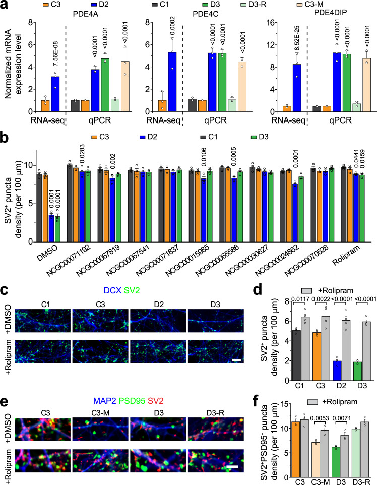
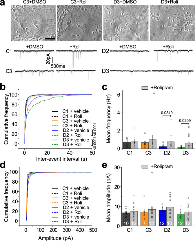
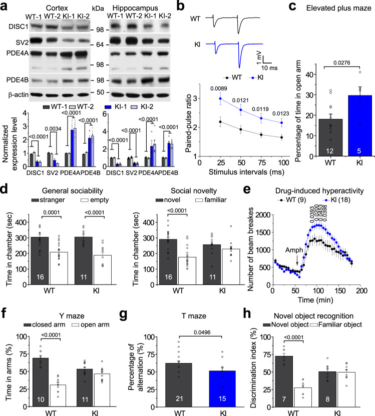
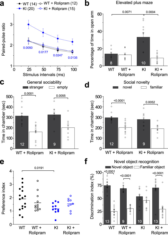
We previously identified a causal link between a rare patient mutation in DISC1 (disrupted-in-schizophrenia 1) and synaptic deficits in cortical neurons differentiated from isogenic patient-derived induced pluripotent stem cells (iPSCs). Here we find that transcripts related to phosphodiesterase 4 (PDE4) signaling are significantly elevated in human cortical neurons differentiated from iPSCs with the DISC1 mutation and that inhibition of PDE4 or activation of the cAMP signaling pathway functionally rescues synaptic deficits. We further generated a knock-in mouse line harboring the same patient mutation in the Disc1 gene. Heterozygous Disc1 mutant mice exhibit elevated levels of PDE4s and synaptic abnormalities in the brain, and social and cognitive behavioral deficits. Pharmacological inhibition of the PDE4 signaling pathway rescues these synaptic, social and cognitive behavioral abnormalities. Our study shows that patient-derived isogenic iPSC and humanized mouse disease models are integral and complementary for translational studies with a better understanding of underlying molecular mechanisms.


Conflict of interest statement

The authors declare no competing interests.

Figures

Fig. 1
Fig. 1. Mechanism-guided pharmacological rescue of synaptic defects of human neurons carrying the DISC1 mutation.
a Summary of q-PCR validation of RNA-seq results of increased mRNA levels of PDE4A, PDE4C, and PDE4DIP in human forebrain neurons carrying the DISC1 mutation (D2 and D3: neurons derived from patient iPSC lines with DISC1 mutation; C1 and C3: neurons derived from control iPSC lines; C3-M: neurons derived from an isogenic knock-in DISC1 mutant line under the background of the C3 control iPSC line; D3-R: neurons derived from an isogenic DISC1 mutant correction line under the background of the D3 patient iPSC line). Values represent mean ± s.d. (n = 3 independent differentiation cultures for qPCR analysis; comparison to C3 using One-way ANOVA with p values indicated). b Effects on the density of SV2+ synaptic puncta in 6-week old forebrain neurons upon treatment with different small molecules. NCGC00071192: 2 µM; NCGC00067819: 2 µM; NCGC00067541: 2 µM; NCGC00071837: 2 µM; NCGC00015985: 10 µM; NCGC00065586: 10 µM; NCGC00030627: 20 µM; NCGC00024862: 20 µM; NCGC00070528: 10 µM; Rolipram: 100 nM. Values represent mean ± s.e.m. (n = 3 independent differentiation cultures; comparison to C1 using One-way ANOVA with p values indicated). cd Representative confocal images of 4-week old neurons from different iPSC lines immunostained with SV2 and DCX upon vehicle (DMSO) or Rolipram (100 nM) treatment. Scale bar, 20 μm (c). Also shown is the quantification of SV2+ puncta density in neurons derived from different iPSC lines with or without Rolipram treatment (d). Values represent mean ± s.e.m. (n = 4 independent differentiation cultures; One-way ANOVA with p values indicated). ef Representative confocal images of 4-week old neurons co-cultured with astrocytes from different iPSC lines immunostained with SV2, PSD95, and MAP2 upon vehicle (DMSO) or Rolipram (100 nM) treatment. Scale bar, 20 μm (e). Also shown is the quantification of SV2+PSD95+ puncta density in neurons derived from different iPSC lines with or without Rolipram treatment (f). Values represent mean ± s.e.m. (n = 3 independent differentiation cultures; One-way ANOVA with p values indicated). Source data are provided as a Source Data file.
Fig. 2
Fig. 2. Functional rescue of synaptic defects of DISC1 mutant neurons.
a Sample images of 6-week-old human neurons differentiated from iPSC lines co-cultured with astrocytes (top) and sample whole-cell voltage-clamp recording traces of excitatory spontaneous synaptic currents (SSCs), with the treatment of vehicle or Rolipram (Roli, 100 nM). Scale bar, 50 μm. The representative images were taken from 3 independent experiments. bc Cumulative distribution plots of SSC inter-event intervals (b, 4 weeks old neurons) and quantification (c, 6 weeks old neurons) showing rescue of reduced SSC frequency of DISC1 mutant neurons after Rolipram treatment. Values in c represent mean ± s.e.m. (n = 3 independent differentiation cultures; cell number 7 to 12 for each condition; Kolmogorov–Smirnov test for b, and One-way ANOVA for c with p values indicated). de Rolipram treatment has no effect on the amplitude of SSCs. Cumulative distribution plots of SSC amplitude (d, 4 weeks old neurons) and quantification (e, 6 weeks old neurons). The same neurons as in bc were examined. Values in e represent mean ± s.e.m. (n = 3 independent differentiation cultures; cell number 7 to 12 for each condition). Source data are provided as a Source Data file.
Fig. 3
Fig. 3. Molecular and behavioral characterization of heterozygous Disc1 KI mice carrying the 4-bp deletion mutation of the Disc1 gene.
a Protein expression levels in cortical and hippocampal lysates of 4-month-old WT and KI mice. KI refers to heterozygous Disc1 mutant mice. Shown are sample western blot images and quantification of protein levels of DISC1, SV2, PDE4A, and PDE4B. Data were normalized to β-actin for sample loading and then to WT in the same blot for comparison. Values represent mean ± s.e.m. (DISC1: n = 6; SV2: n = 2; PDE4A: n = 4; PDE4B: n = 6 independent experiments for cortex; DISC1: n = 5; SV2: n = 8; PDE4A: n = 6; PDE4B: n = 5 independent experiments for hippocampus; One-way ANOVA with p values indicated). b Impaired paired-pulse ratio in Disc1 KI mice. The electrophysiological response of CA1 neurons to paired pulses of stimulation of CA3 neurons from acutely prepared hippocampal slices. The paired pulses were separated by intervals of 25, 50, 75, and 100 ms. Values represent mean ± s.e.m. (n = 14 for WT and n = 20 for KI; One-way ANOVA with p values indicated). c Basal anxiety of Disc1 KI mice in the elevated plus-maze test. The percentage of time spent in the open arm over total time was calculated. Values represent mean ± s.e.m. (n = 12 for WT and n = 5 for KI, 13-week-old male littermates; One-way ANOVA with p values indicated). d Three-chamber social preference test showing impaired social novelty recognition of Disc1 KI mice (right), but normal general sociability (left). Time spent in each chamber was calculated. Values represent mean ± s.e.m. (n = 16 for WT and n = 11 for KI, 16-week-old male littermates; One-way ANOVA with p values indicated). e Disc1 KI mice showed a higher level of D-amphetamine (Amph)-induced hyperactivity. Locomotor activity was measured 60 min before and 120 min immediately after Amph treatment. A total number of beam breaks was measured. Data are shown in 5 min intervals. Values represent mean ± s.e.m. (n = 9 for WT and n = 18 for KI, 16-week-old male littermates; One-way ANOVA with p values indicated). f Impaired spatial working memory of Disc1 KI mice in the Y-maze task. The time spent in each arm during the 5 min test was shown. Values represent mean ± s.e.m. (n = 10 for WT and n = 11 for KI, 14-week-old male littermates; One-way ANOVA with p values indicated). g Impaired spontaneous alternation of Disc1 KI mice in the T-maze task. The percentage of alternations (alternated arm entry on two consecutive trials) was scored for 15 trials. Values represent mean ± s.e.m. (n = 21 for WT and n = 15 for KI, 18-week-old male littermates; One-way ANOVA with p values indicated). h Decrease of discriminative activity of Disc1 KI mice in the novel object recognition test. KI mice showed no preference for the novel object over the familiar object. Values represent mean ± s.e.m. (n = 7 for WT and n = 8 for KI, 15-week-old male littermates; One-way ANOVA with p values indicated). Source data are provided as a Source Data file.
Fig. 4
Fig. 4. Synaptic and behavioral impairments in Disc1 KI mice rescued by Rolipram treatment.
a Rolipram restored synaptic transmission deficits in the hippocampus of Disc1 KI mice. Rolipram (0.1 mg/kg) or vehicle was injected 30 min before sacrifice and was present in the bath solution (0.7 μM) throughout the recording. Values represent mean ± s.e.m.; (n is indicated in the plot; comparison between KI and KI + Rolipram using One-way ANOVA with p values indicated). The WT and KI without Rolipram treatment from Fig. 3b were replotted here for comparison. b Rolipram normalized the anxiety level of Disc1 KI mice in the elevated plus maze test. Values represent mean ± s.e.m. (n = 8 for WT and n = 10 for KI, 13-week-old male littermates; One-way ANOVA with p values indicated). ce Rolipram rescued the social behavioral deficits of Disc1 KI mice in the 3 chambers social interaction test. There was no effect of Rolipram on general sociability (c), but Rolipram treatment restored the social novelty recognition of KI mice (d). Shown in e is a summary of the calculated preference index for novel and familiar mice with or without Rolipram treatment. Values represent mean ± s.e.m. (n = 16 for WT, n = 13 for WT + Rolipram, = 11 for KI, and n = 10 for KI + Rolipram; 16-week-old male littermates; One-way ANOVA with p values indicated). f Rolipram rescued the cognitive-behavioral deficits of Disc1 KI mice in the novel object recognition test. Values represent mean ± s.e.m. (n is indicated in the plot, 15-week-old male littermates; One-way ANOVA with p values indicated). Source data are provided as a Source Data file.

Similar articles

Cited by

References

    1. Coyle JT, Ruzicka WB, Balu DT. Fifty years of research on schizophrenia: the ascendance of the glutamatergic synapse. Am. J. Psychiatry. 2020;177:1119–1128. - PMC - PubMed
    1. Hall J, Trent S, Thomas KL, O’Donovan MC, Owen MJ. Genetic risk for schizophrenia: convergence on synaptic pathways involved in plasticity. Biol. Psychiatry. 2015;77:52–58. - PubMed
    1. Brennand KJ, Simone A, Tran N, Gage FH. Modeling psychiatric disorders at the cellular and network levels. Mol. Psychiatry. 2012;17:1239–1253. - PMC - PubMed
    1. Bellin M, Marchetto MC, Gage FH, Mummery CL. Induced pluripotent stem cells: the new patient? Nat. Rev. Mol. Cell Biol. 2013;13:713–726. - PubMed
    1. Soliman MA, Aboharb F, Zeltner N, Studer L. Pluripotent stem cells in neuropsychiatric disorders. Mol. Psychiatry. 2017;22:1241–1249. - PMC - PubMed

Publication types

MeSH terms