

Giant Intraparenchymal Meningioma in a Female Child: Case Report and Literature Review
- PMID: 33658857
- PMCID: PMC7920497
- DOI: 10.2147/CMAR.S294224
Giant Intraparenchymal Meningioma in a Female Child: Case Report and Literature Review
Abstract
Background: Intraparenchymal meningiomas without dural attachment are extremely rare, especially in female children. To our knowledge, fibrous intraparenchymal meningioma located in the temporal lobe has never been reported in female children. The significance in the differential diagnosis of lesions in the temporal lobe should be emphasized.
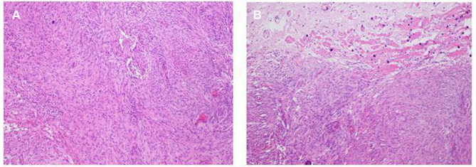
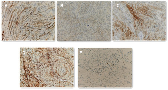
Case presentation: A 12-year-old girl was admitted to our hospital, complaining of recurrent generalized seizures for 2 months. Magnetic resonance imaging demonstrated a solid lesion located in the temporal lobe. The lesion underwent gross total resection. Histopathological examination indicated that the lesion was a fibrous meningioma. Postoperative rehabilitation was uneventful.
Conclusion: This case report presents an extremely unusual intraparenchymal fibrous meningioma of the temporal lobe with peritumoral edema and reviewed 21 intraparenchymal meningioma cases in children and to discuss the clinical presentation and treatment, differential diagnosis, and radiological features.
Keywords: fibrous; intraparenchymal; meningioma; temporal lobe.
© 2021 Guo et al.
Conflict of interest statement
The authors declare that they have no competing interests.
Figures

Similar articles
-
Intraparenchymal Atypical Meningioma in Basal Ganglia Region in a Child: Case Report and Literature Review.J Korean Neurosurg Soc. 2018 Jan;61(1):120-126. doi: 10.3340/jkns.2015.0609.001. Epub 2017 Dec 29. J Korean Neurosurg Soc. 2018. PMID: 29354244 Free PMC article.
-
Intraparenchymal atypical meningioma in the posterior fossa: a case report and literature review.Br J Neurosurg. 2023 Oct;37(5):1167-1170. doi: 10.1080/02688697.2021.1884651. Epub 2021 Feb 17. Br J Neurosurg. 2023. PMID: 33595378 Review.
-
Intraparenchymal meningioma in a child. Case report and review of the literature.J Neurosurg. 2004 Aug;101(1 Suppl):112-5. doi: 10.3171/ped.2004.101.2.0112. J Neurosurg. 2004. PMID: 16206982
-
Intraparenchymal meningioma mimicking cavernous malformation: a case report and review of the literature.J Med Case Rep. 2014 Dec 29;8:467. doi: 10.1186/1752-1947-8-467. J Med Case Rep. 2014. PMID: 25547419 Free PMC article. Review.
-
Intraparenchymal Meningioma: Clinical, Radiologic, and Histologic Review.World Neurosurg. 2016 Aug;92:23-30. doi: 10.1016/j.wneu.2016.04.098. Epub 2016 May 4. World Neurosurg. 2016. PMID: 27155381 Review.
Cited by
-
Interhemispheric Pediatric Meningioma, YAP1 Fusion-Positive.Diagnostics (Basel). 2022 Sep 29;12(10):2367. doi: 10.3390/diagnostics12102367. Diagnostics (Basel). 2022. PMID: 36292056 Free PMC article.
-
Pediatric Meningiomas: Current Insights on Pathogenesis and Management.Acta Neurochir Suppl. 2023;135:69-74. doi: 10.1007/978-3-031-36084-8_12. Acta Neurochir Suppl. 2023. PMID: 38153451 Review.
-
Imaging features of pediatric meningiomas: emphasis on unusual locations.Childs Nerv Syst. 2024 Dec;40(12):3933-3942. doi: 10.1007/s00381-024-06525-2. Epub 2024 Jul 10. Childs Nerv Syst. 2024. PMID: 38985317
-
Incidental pediatric intraparenchymal meningioma: illustrative case.J Neurosurg Case Lessons. 2025 May 19;9(20):CASE24611. doi: 10.3171/CASE24611. Print 2025 May 19. J Neurosurg Case Lessons. 2025. PMID: 40388891 Free PMC article.
References
Publication types
LinkOut - more resources
Full Text Sources
Other Literature Sources
