. 2021 Feb 15:15:633018.
doi: 10.3389/fnins.2021.633018. eCollection 2021.

Functional and Neurochemical Identification of Ghrelin Receptor (GHSR)-Expressing Cells of the Lateral Parabrachial Nucleus in Mice

Affiliations

Functional and Neurochemical Identification of Ghrelin Receptor (GHSR)-Expressing Cells of the Lateral Parabrachial Nucleus in Mice

Marie V Le May et al. Front Neurosci. .

Abstract

The lateral parabrachial nucleus (lPBN), located in the pons, is a well-recognized anorexigenic center harboring, amongst others, the calcitonin gene-related peptide (CGRP)-expressing neurons that play a key role. The receptor for the orexigenic hormone ghrelin (the growth hormone secretagogue receptor, GHSR) is also abundantly expressed in the lPBN and ghrelin delivery to this site has recently been shown to increase food intake and alter food choice. Here we sought to explore whether GHSR-expressing cells in the lPBN (GHSR lPBN cells) contribute to feeding control, food choice and body weight gain in mice offered an obesogenic diet, involving studies in which GHSR lPBN cells were silenced. We also explored the neurochemical identity of GHSR lPBN cells. To silence GHSR lPBN cells, Ghsr-IRES-Cre male mice were bilaterally injected intra-lPBN with a Cre-dependent viral vector expressing tetanus toxin-light chain. Unlike control wild-type littermates that significantly increased in body weight on the obesogenic diet (i.e., high-fat high-sugar free choice diet comprising chow, lard and 9% sucrose solution), the heterozygous mice with silenced GHSR lPBN cells were resistant to diet-induced weight gain with significantly lower food intake and fat weight. The lean phenotype appeared to result from a decreased food intake compared to controls and caloric efficiency was unaltered. Additionally, silencing the GHSR lPBN cells altered food choice, significantly reducing palatable food consumption. RNAscope and immunohistochemical studies of the lPBN revealed considerable co-expression of GHSR with glutamate and pituitary adenylate cyclase-activating peptide (PACAP), and much less with neurotensin, substance P and CGRP. Thus, the GHSR lPBN cells are important for diet-induced weight gain and adiposity, as well as in the regulation of food intake and food choice. Most GHSR lPBN cells were found to be glutamatergic and the majority (76%) do not belong to the well-characterized anorexigenic CGRP cell population.

Keywords: CGRP; GHSR; PACAP; body weight; food intake and food choice; ghrelin; glutamate; parabrachial nucleus.


Conflict of interest statement

The authors declare that the research was conducted in the absence of any commercial or financial relationships that could be construed as a potential conflict of interest.

Figures

FIGURE 1
FIGURE 1
Effect of GHSRlPBN cells Tetox-silencing on body weight, epididymal fat pads and caloric efficiency. (A) Body weight at the start and on day 23 (D23) of access to high-fat, high-sugar (HFHS) free choice diet for both the control group (lighter grey) and the GHSR-silenced group (darker grey). (B) Weight of bilateral epididymal fat pads at the end of the experiment for both groups. (C) Caloric efficiency of both groups on day 23 of HFHS free choice diet. (D) Evolution of % body weight gain of both groups on HFHS free choice diet over time (body weight at start of HFHS diet = 100%). Data shown as mean ± SEM. p < 0.05 and ∗∗p ≤ 0.01.
FIGURE 2
FIGURE 2
Effect of GHSRlPBN cells Tetox-silencing on food choice and energy intake. (A) Average daily energy intake from chow, lard, 9% sucrose solution, lard and 9% sucrose solution together, total energy intake (kcal) and water intake (in ml) calculated over 1 week. (B) Evolution of total energy intake of both groups on HFHS free choice diet over time. Data shown as mean ± SEM. p < 0.05, ∗∗p ≤ 0.01, ∗∗∗p ≤ 0.001.
FIGURE 3
FIGURE 3
Co-expression of glutamate and pituitary adenylate cyclase-activating peptide (PACAP) with GHSR in the lPBN of mice. (A,B) Representative images of the lPBN with RNAscope for glutamate transporter (Slc17a6) and Ghsr mRNAs (with DAPI) and for the PACAP-coding gene (Adcyap1) and Ghsr mRNAs (with DAPI), respectively. (C–E) Magnifications of the indicated part of A with the signals from Slc17a6 and Ghsr mRNAs separated and merged. (G–I) Magnifications of the indicated part of B with the signals from Adcyap1 and Ghsr mRNAs separated and merged. (F,J) Quantification of co-expression of Ghsr mRNA with Slc17a6 mRNA (F) and with Adcyap1 mRNA (J) in the lPBN of 4 mice (5 lPBN sections per mouse). MoleculeXMoleculeY indicates the percentage of MoleculeY-expressing cells co-expressing MoleculeX. Scale bar = 50 μm. scp: superior cerebellar peduncle.
FIGURE 4
FIGURE 4
Co-expression of neurotensin and substance P with GHSR in the lPBN of mice. (A,B) Representative images of the lPBN with RNAscope for neurotensin (Nts) and Ghsr mRNAs (with DAPI) and for the substance P-coding gene (Tac1) and Ghsr mRNAs (with DAPI), respectively. (C–E) Magnifications of the indicated part of A with the signals from neurotensin and Ghsr mRNAs separated and merged. (G–I) Magnifications of the indicated part of B with the signals from Tac1 and Ghsr mRNAs separated and merged. (F,J) Quantification of co-expression of Ghsr mRNA with neurotensin mRNA (Nts, F) and with Tac1 mRNA (J) in the lPBN of 4 mice (4–5 lPBN sections per mouse). MoleculeXMoleculeY indicates the percentage of MoleculeY-expressing cells co-expressing MoleculeX. Scale bar = 50 μm. scp: superior cerebellar peduncle.
FIGURE 5
FIGURE 5
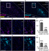
Co-expression of calcitonin gene-related peptide (CGRP) with GHSR in the lPBN of mice. (A) Representative image of the lPBN with immunohistochemical staining for CGRP and endogenous expression of ZsGreen in GHSR-expressing cells of Ghsr-IRES-Cre heterozygous mice on a ZsGreen background (with DAPI). (B) Quantification of co-expression of GHSR (visualized as ZsGreen) and CGRP in the lPBN of 5 mice (2 lPBN sections per mouse). MoleculeXMoleculeY indicates the percentage of MoleculeY-expressing cells co-expressing MoleculeX. (C–E) Magnifications of the indicated part of A with the signals from CGRP and ZsGreen separated and merged. Filled arrow heads: ZsGreen-expressing cells, hollow arrow heads: CGRP-positive cells, filled arrow: cell expressing both ZsGreen and CGRP. Scale bar = 50 μm. scp: superior cerebellar peduncle.

Similar articles

Cited by

References

    1. Alhadeff A. L., Baird J. P., Swick J. C., Hayes M. R., Grill H. J. (2014a). Glucagon-like Peptide-1 receptor signaling in the lateral parabrachial nucleus contributes to the control of food intake and motivation to feed. Neuropsychopharmacology 39 2233–2243. 10.1038/npp.2014.74 - DOI - PMC - PubMed
    1. Alhadeff A. L., Hayes M. R., Grill H. J. (2014b). Leptin receptor signaling in the lateral parabrachial nucleus contributes to the control of food intake. Am. J. Physiol. Regul. Integr. Comp. Physiol. 307 R1338–R1344. - PMC - PubMed
    1. Alhadeff A. L., Golub D., Hayes M. R., Grill H. J. (2015). Peptide YY signaling in the lateral parabrachial nucleus increases food intake through the Y1 receptor. Am. J. Physiol. Endocrinol. Metab. 309 E759–E766. - PMC - PubMed
    1. Bake T., Le May M. V., Edvardsson C. E., Vogel H., Bergström U., Albers M. N., et al. (2020). Ghrelin receptor stimulation of the lateral parabrachial nucleus in rats increases food intake but not food motivation. Obesity (Silver Spring). 28 1503–1511. 10.1002/oby.22875 - DOI - PubMed
    1. Bernard J. F., Alden M., Besson J. M. (1993). The organization of the efferent projections from the pontine parabrachial area to the amygdaloid complex: a Phaseolus vulgaris leucoagglutinin (PHA-L) study in the rat. J. Comp. Neurol. 329 201–229. 10.1002/cne.903290205 - DOI - PubMed

LinkOut - more resources