Case Reports
. 2021 Feb 11;16(4):934-937.
doi: 10.1016/j.radcr.2021.01.061. eCollection 2021 Apr.

Inferior vena cava filter fracture and migration to the pulmonary artery

Affiliations
Case Reports

Inferior vena cava filter fracture and migration to the pulmonary artery

Jaehyung Kim. Radiol Case Rep. .

Erratum in

Abstract

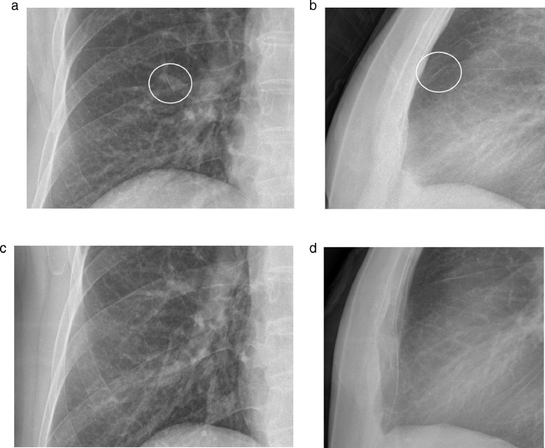
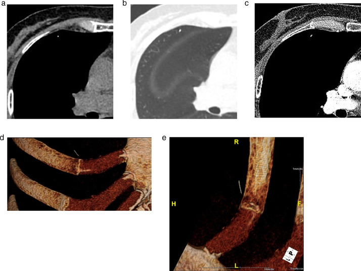
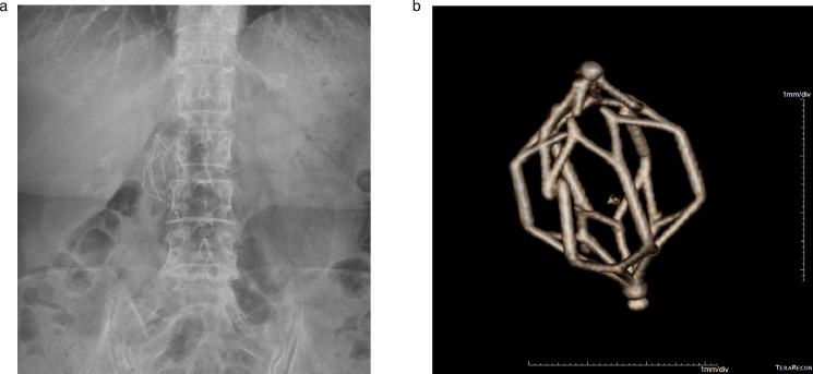
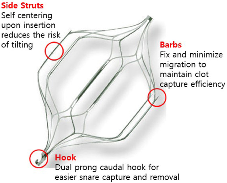
Inferior vena cava (IVC) filters provide a safe and effective method for protecting against pulmonary embolisms in patients for whom standard anticoagulation therapy for acute deep vein thrombosis is contraindicated. Common complications of IVC filter placement include erosion through the wall of the vena cava, visceral perforation, and filter thrombosis, obstruction, and migration. In this report, we describe the case of a 60-year-old woman who presented with an IVC filter fracture and subsequent migration of the filter to the lung detected via chest radiography.

Keywords: Filter fracture; Filter strut migration to pulmonary artery; IVC filter complication.


Figures

Fig 1 –
Fig. 1
Plain chest PA (A) and lateral (B) radiographs 8 years after filter placement showing a single linear opacity in the right upper lung field (white circle). Plain chest PA (C) and lateral (D) radiographs 6 years after filter placement showing no abnormal finding in the same region.
Fig 2 –
Fig. 2
Precontrast (A), lung window (B), and postcontrast (C) axial CT scans of the chest demonstrating the fractured and migrated IVC filter strut as a bright radiopaque structure within a right pulmonary artery branch. Volume rendering images (D,E) showing a linear radiopaque structure within a right pulmonary artery branch.
Fig 3 –
Fig. 3
Plain abdomen radiograph (A) and the 3D reconstructed image (B) showing the fractured OptEase IVC filter in the IVC.
Fig 4 –
Fig. 4
Side view of the OptEase filter demonstrating a symmetrical double-basket design, which consist with the six straight struts engaged the proximal and distal baskets and anchoring barbs at the upper pole of each of the six straight struts.

References

    1. Grewal S, Chamarthy MR, Kalva SP. Complications of inferior vena cava filters. Cardiovasc Diagn Ther. 2016;6(6):632–641. doi: 10.21037/cdt.2016.09.08. - DOI - PMC - PubMed
    1. Vossen JA, Thawait SK, Golia JS, Chamarthy M, Cholewczynski W, Velasco N. Recurrent fracture of a recovery inferior vena cava filter with pulmonary migration. Yale j Biol Med. 2012;85(2):255–259. - PMC - PubMed
    1. Mitsunaga MM, Yoon H. Fracture rate and serious complications of vena cava filters. Open J Rad. 2013;3(2):85–90. doi: 10.4236/ojrad.2013.32013. - DOI
    1. Ziegler JW, Dietrich GJ, Cohen SA, Sterling K, Duncan J, Samotowka M. PROOF trial: protection from pulmonary embolism with the OptEase filter. J Vasc Interv Radiol. 2008;19(8):1165–1170. doi: 10.1016/j.jvir.2008.04.020. - DOI - PubMed
    1. Rogers NA, Nguyen L, Minniefield NE, Jessen ME, de Lemos JA. Fracture and embolization of an inferior vena cava filter strut leading to cardiac tamponade. Circulation. 2009;119(18):2535–2536. doi: 10.1161/CIRCULATIONAHA.108.817247. - DOI - PubMed

Publication types

LinkOut - more resources