. 2021 Feb 15:10:614096.
doi: 10.3389/fonc.2020.614096. eCollection 2020.

Three-Dimensional Ex Vivo Culture for Drug Responses of Patient-Derived Gastric Cancer Tissue

Affiliations

Three-Dimensional Ex Vivo Culture for Drug Responses of Patient-Derived Gastric Cancer Tissue

Sian Chen et al. Front Oncol. .

Abstract

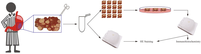
Gastric cancer (GC) is one of the most common malignancies with high mortality and substantial morbidity. Although the traditional treatment strategies for GC revolve around surgery, radiotherapy, and chemotherapy, none have been able to optimally treat most affected patients. To improve clinical outcomes and overcome potential GC resistance, we established a three-dimensional (3D) culturing platform that accurately predicts drug responses in a time- and cost-effective manner. We collected tumor tissues from patients following surgeries and cultured them for 3 days using our protocol. We first evaluated cell proliferation, viability, and apoptosis using the following markers: Ki67 and cleaved caspase 3 (Cas3). We demonstrated that cell viability was maintained for 72 h in culture and that the tumor microenvironments and vascular integrities of the tissues were intact throughout the culture period. We then administered chemotherapeutics to assess drug responses and found differential sensitivity across different patient-derived tissues, enabling us to determine individualized medication plans. Overall, our study validated this rapid, cost-effective, scalable, and reproducible protocol for GC tissue culture that can be employed for drug response assessments. Our 3D culture platform paves a new way for personalized medication in GC and other tumors and can greatly impact future oncological research.

Keywords: apoptosis; chemotherapy effectiveness; ex vivo tumor tissue culture; gastric cancer; proliferation.


Conflict of interest statement

The authors declare that the research was conducted in the absence of any commercial or financial relationships that could be construed as a potential conflict of interest.

Figures

Figure 1
Figure 1
Tumor tissue collection, preparation, and culture.
Figure 2
Figure 2
Cell diversity in GC tissue culture. (A) After 3 days culture, the patient-derived GC tissues (Case3) were stained with α-SMA, fibronectin, collagen, and counterstained with DAPI. Scale bars are all 100 μm. (B–D) Quantitative analysis of α-SMA, fibronectin, and collagen expression during the 3 days culture.
Figure 3
Figure 3
GC Cell Proliferation and Apoptosis during the culture. (A) Hematoxylin staining of Case13 before and after 3 days of culture. Cas3 staining of Case15 before and after 3 days of culture. Ki67 staining of Case16 before and after 3 days of culture, scale bar, 100 μm. (B) Quantitative analysis of hematoxylin in 20 cases of GC tissues. (C) Quantitative analysis of Cas3 in 20 cases of GC tissues. (D) Quantitative analysis of Ki67 in 20 cases of GC tissues.
Figure 4
Figure 4
EdU incorporation and hypoxia of GC cell during the culture. (A) EdU incorporation of Case29 before and after 3 days of culture, scale bar, 50 μm, and HIF-1α staining of Case30 before and after 3 days of culture, scale bar, 50 μm. (B) Quantitative analysis of EdU in Case29. (C) Quantitative analysis of HIF-1α in Case30.
Figure 5
Figure 5
Cancer cellular and structural integrities within GC tissue. (A) Keratin20 staining of Case1 before and after 3 days of culture, scale bar, 100 μm. CD133 staining of Case4 before and after 3 days of culture, scale bar, 100 μm. (B) Case13 was intestinal type carcinoma and Case17 was diffuse gastric carcinoma, and the H&E staining at different time points were laid out. Compared with results before culture (day0), the cancer cell distributions and structure integrities were not changed a lot after 3 days of culture. All images are the same magnification, scale bar, 100 μm. (C) Keratin20 quantitative analysis of Case1-Case5 before and after 3 days of culture. (D) CD133 quantitative analysis of Case1-Case5 before and after 3 days of culture.
Figure 6
Figure 6
Results of drug response tests to oxaliplatin based on 3D GC tissue culture. (A) The tumor tissues of Case22 and Case24 were stained with Cas3 after 3 days of culture and oxaliplatin treatment. (B) The tumor tissues of Case22 and Case24 were stained with Ki67 after 3 days of culture and oxaliplatin treatment. All images are the same magnification, scale bar, 100 μm. (C, D) Quantitative analysis of Ki67 and Cas3 in tissues of Case21-25. (E) The tumor tissues of Case22 and Case24 were stained with p53 after 3 days of culture and oxaliplatin treatment. All images are the same magnification, scale bar, 100 μm. (F) Quantitative analysis of p53 in tissues of Case22 and Case24.
Figure 7
Figure 7
Results of drug response tests to 5-FU based on 3D GC tissue culture. (A) The tumor tissues of Case26, Case27, and Case28 were stained with Cas3 after 3 days of culture and 5-FU treatment. (B) The tumor tissues of Case26, Case27, and Case28 were stained with Ki67 after 3 days of culture and 5-FU treatment. All images are the same magnification, scale bar, 100 μm. (C, D) Quantitative analysis of Ki67 and Cas3 in tissues of Case26-28.

References

    1. Bray F, Ferlay J, Soerjomataram I, Siegel RL, Torre LA, Jemal A. Global cancer statistics 2018: GLOBOCAN estimates of incidence and mortality worldwide for 36 cancers in 185 countries. CA Cancer J Clin (2018) 68(6):394–424. 10.3322/caac.21492 - DOI - PubMed
    1. Jackson SE, Sedikides GX, Okecha G, Wills MR. Generation, maintenance and tissue distribution of T cell responses to human cytomegalovirus in lytic and latent infection. Med Microbiol Immunol (2019) 208(3-4):375–89. 10.1007/s00430-019-00598-6 - DOI - PMC - PubMed
    1. Yang L, Zheng R, Wang N, Yuan Y, Liu S, Li H, et al. . Incidence and mortality of stomach cancer in China, 2014. Chin J Cancer Res (2018) 30(3):291–8. 10.21147/j.issn.1000-9604.2018.03.01 - DOI - PMC - PubMed
    1. China NHCotPsRo . Standards for gastric cancer diagnosis and treatment (2018 edition). Multi Cancer Treat (2019) 5(1):55–82. 10.12151/JMCM.2019.01-08 - DOI
    1. Balakrishnan M, George R, Sharma A, Graham DY. Changing trends in stomach cancer throughout the world. Curr Gastroenterol Rep (2017) 19(8):36. 10.1007/s11894-017-0575-8 - DOI - PMC - PubMed