

Analysis of Generalized Fibrosis in Mouse Tissue Sections with Masson's Trichrome Staining
- PMID: 33659302
- PMCID: PMC7842772
- DOI: 10.21769/BioProtoc.3629
Analysis of Generalized Fibrosis in Mouse Tissue Sections with Masson's Trichrome Staining
Abstract
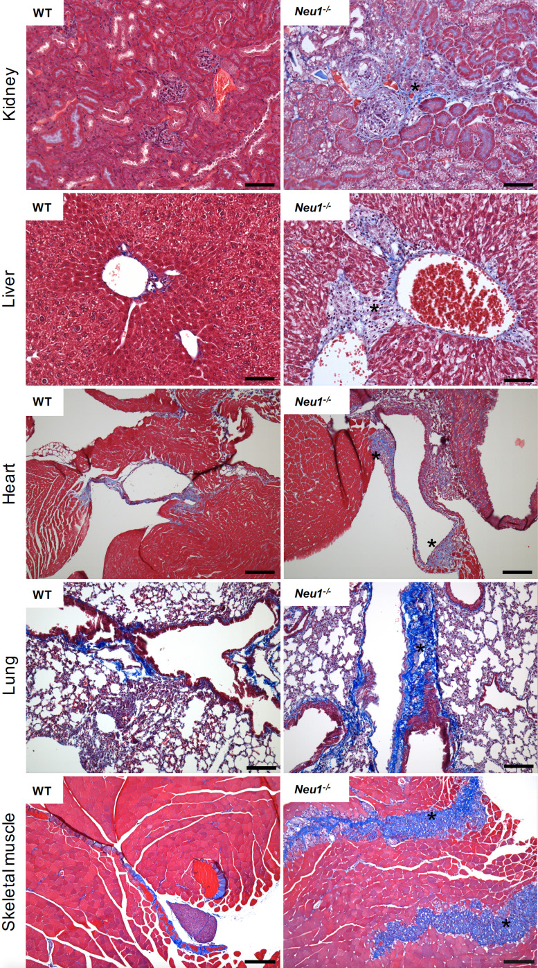
Expansion of fibrous connective tissue and abnormal deposition of extracellular matrix (ECM) are at the basis of many fibrotic diseases. Fibrosis can occur in response to both physiological and pathological cues, including wound healing, tissue remodeling/repair and inflammation. Chronic fibrosis can lead to severe tissue damage, organ failure and death. Assessing the extent of organ fibrosis is crucial for accurate diagnosis of this condition. The use of Masson's trichrome staining of tissue sections from skeletal muscle is a fast method for detection of morphological alterations indicative of a fibrotic phenotype in this organ. This staining method detects the extent of collagen fibers deposition and, because it employs the combination of three dyes, can also distinguish muscle fibers (red), from collagen (blue) and nuclei (black), simultaneously.
Keywords: Collagen; Fibroblasts; Fibrosis; Masson’s Trichrome; Skeletal muscle; Tissue section.
Copyright © 2020 The Authors; exclusive licensee Bio-protocol LLC.
Conflict of interest statement
Competing interestsThe authors have no conflict of interest or competing interests to declare.
Figures
References
-
- Carson F.(1990). Histotechnology A Self-Instructional Text. 1990, 1st Ed, pp142-143, ASCP, Ill
-
- de Geest N., Bonten E., Mann L., de Sousa-Hitzler J., Hahn C. and d'Azzo A.(2002). Systemic and neurologic abnormalities distinguish the lysosomal disorders sialidosis and galactosialidosis in mice. Hum Mol Genet 11(12): 1455-1464. - PubMed
-
- Hinderer S. and Schenke-Layland K.(2019). Cardiac fibrosis- A short review of causes and therapeutic strategies. Adv Drug Deliv Rev 146: 77-82. - PubMed
Grants and funding
LinkOut - more resources
Full Text Sources
