

Mouse Adipose Tissue Protein Extraction
- PMID: 33659303
- PMCID: PMC7842653
- DOI: 10.21769/BioProtoc.3631
Mouse Adipose Tissue Protein Extraction
Abstract
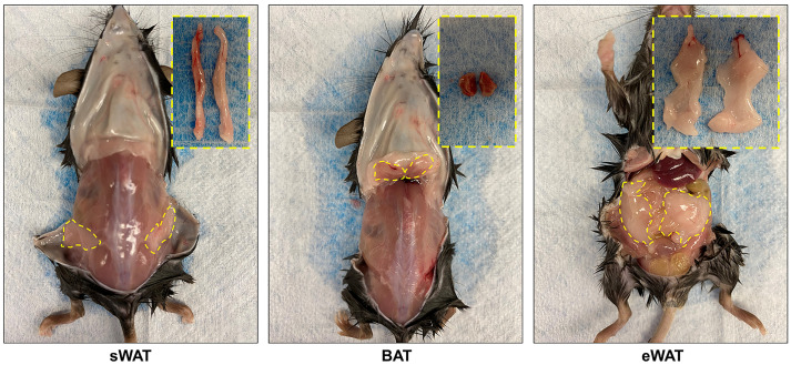
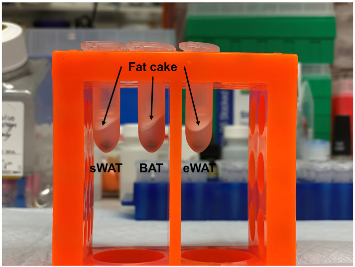
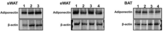
As obesity becomes a global epidemic, the metabolism research field is increasingly focusing on studying the physiological and pathological roles of adipose tissues (AT). However, extracting proteins from AT is challenging due to abundant fat content of intracellular lipid droplets. Several commercial kits for extraction of AT proteins are available, as are protocols (such as the RELi protocol as well as other protein precipitation protocols). The protocols have been introduced to improve the quality and yield of extractions, but these methods either increase the cost or involve multiple steps. Herein, we describe a detailed protocol for mouse AT protein extractions based on our daily laboratory practice. This protocol requires only very common reagents and instruments, and can be completed in 90-120 min and provides good recovery of total protein content. Thus, this protocol is an economically attractive, time-saving and efficient way to extract proteins from the AT.
Keywords: Adipose Tissue; Lipid Contamination; Metabolism; Mouse; Obesity; Protein; Time and Cost Effective.
©Copyright An and Scherer.
Conflict of interest statement
Competing interestsThe authors declare no competing interests.
Figures
References
-
- An Y. A., Crewe C., Asterholm I. W., Sun K., Chen S., Zhang F., Shao M., Funcke J.-B., Zhang Z., Straub L., Yoshino J., Klein S., Kusminski C. M. and Scherer P. E.(2019). Dysregulation of amyloid precursor protein impairs adipose tissue mitochondrial function and promotes obesity. Nature Metabolism 1(12): 1243-1257. - PMC - PubMed
-
- Benabdelkamel H., Masood A., Alanazi I. O. and Alfadda A. A.(2018). Comparison of protein precipitation methods from adipose tissue using difference gel electrophoresis. Electrophoresis. - PubMed
Grants and funding
LinkOut - more resources
Full Text Sources
Research Materials
