

Isolation and ex vivo Expansion of Human Limbal Epithelial Progenitor Cells
- PMID: 33659413
- PMCID: PMC7842399
- DOI: 10.21769/BioProtoc.3754
Isolation and ex vivo Expansion of Human Limbal Epithelial Progenitor Cells
Abstract
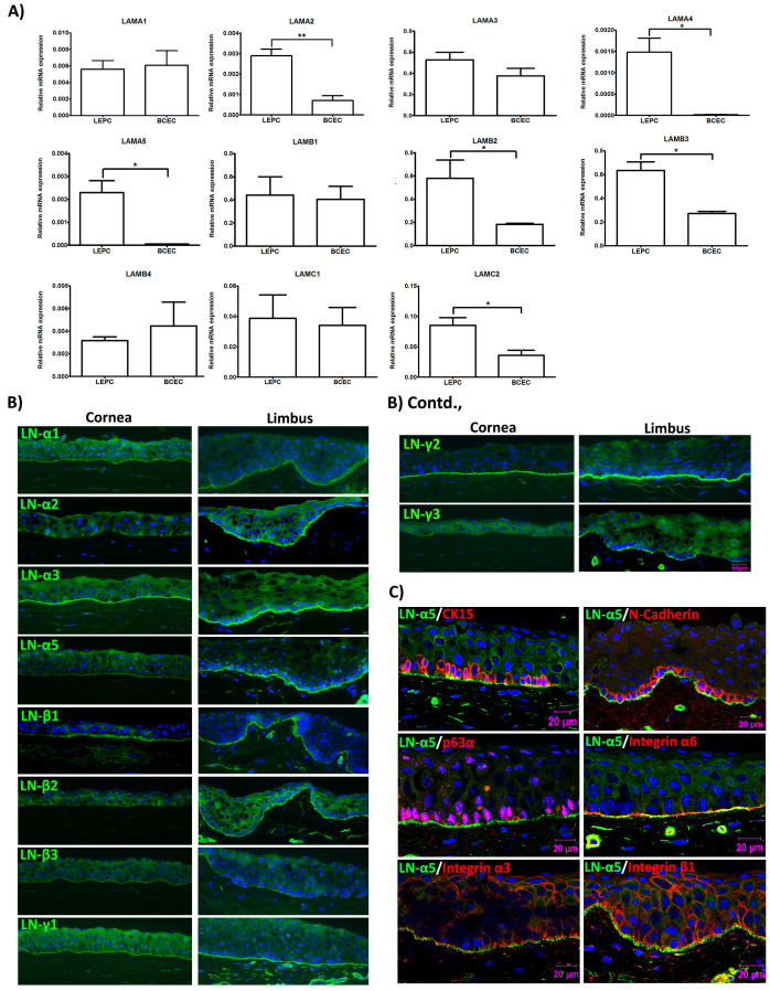
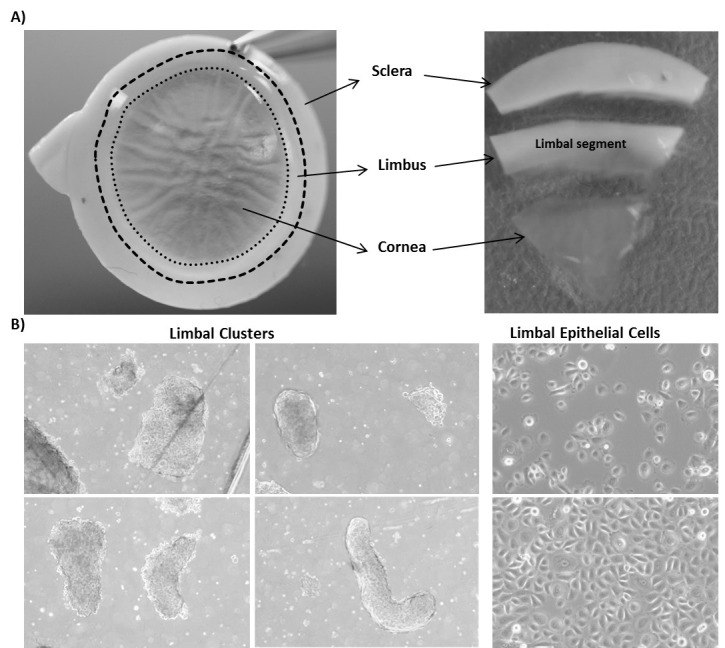
Limbal stem cell transplantation has been used successfully to treat patients with limbal stem cell deficiency all over the world. However, long term clinical results often proved less satisfactory due to the low quality of the graft or inadequate properties of transplanted cells. To enhance the ex vivo expansion of human limbal epithelial stem or progenitor cells (LEPC) by preserving stem cell phenotype and to improve subsequent transplantation efficiency, cell-matrix interactions ex vivo should mimic the condition in vivo. The laminin isoforms preferentially expressed in the limbal niche can be used as a culture matrix for epithelial tissue engineering. We recently published the expansion of LEPC on various laminin isoforms and observed that laminin alpha 5-derived matrices support the efficient expansion of LEPC compared to tissue culture plates and other laminin isoforms by preserving stem/progenitor cell phenotype. Here, we describe an optimized protocol for the isolation of LEPC from cadaveric corneal limbal tissue by collagenase digestion and efficient expansion of LEPC using recombinant human laminin-511 E8 fragment (LN-511E8) as culture substrate.
Keywords: Cornea; Expansion; Isolation; Laminin; Limbal epithelial progenitor cells; Limbal stem cells.
Copyright © 2020 The Authors; exclusive licensee Bio-protocol LLC.
Conflict of interest statement
Competing interestsThe authors declare that they have no competing interests.
Figures
Similar articles
-
Efficient Isolation and Functional Characterization of Niche Cells from Human Corneal Limbus.Int J Mol Sci. 2022 Mar 2;23(5):2750. doi: 10.3390/ijms23052750. Int J Mol Sci. 2022. PMID: 35269891 Free PMC article.
-
Isolation and ex vivo Expansion of Limbal Mesenchymal Stromal Cells.Bio Protoc. 2022 Jul 20;12(14):e4471. doi: 10.21769/BioProtoc.4471. eCollection 2022 Jul 20. Bio Protoc. 2022. PMID: 35978577 Free PMC article.
-
Laminin-511 and -521-based matrices for efficient ex vivo-expansion of human limbal epithelial progenitor cells.Sci Rep. 2017 Jul 11;7(1):5152. doi: 10.1038/s41598-017-04916-x. Sci Rep. 2017. PMID: 28698551 Free PMC article.
-
Characterization, isolation, expansion and clinical therapy of human corneal epithelial stem/progenitor cells.J Stem Cells. 2014;9(2):79-91. J Stem Cells. 2014. PMID: 25158157 Review.
-
Ex vivo expansion of corneal limbal epithelial/stem cells for corneal surface reconstruction.Eur J Ophthalmol. 2003 Jul;13(6):515-24. doi: 10.1177/112067210301300602. Eur J Ophthalmol. 2003. PMID: 12948308 Review.
Cited by
-
Efficient Isolation and Functional Characterization of Niche Cells from Human Corneal Limbus.Int J Mol Sci. 2022 Mar 2;23(5):2750. doi: 10.3390/ijms23052750. Int J Mol Sci. 2022. PMID: 35269891 Free PMC article.
-
Efficient Isolation and Expansion of Limbal Melanocytes for Tissue Engineering.Int J Mol Sci. 2023 Apr 25;24(9):7827. doi: 10.3390/ijms24097827. Int J Mol Sci. 2023. PMID: 37175529 Free PMC article.
-
Isolation and ex vivo Expansion of Limbal Mesenchymal Stromal Cells.Bio Protoc. 2022 Jul 20;12(14):e4471. doi: 10.21769/BioProtoc.4471. eCollection 2022 Jul 20. Bio Protoc. 2022. PMID: 35978577 Free PMC article.
References
-
- Cotsarelis G., Cheng S. Z., Dong G., Sun T. T. and Lavker R. M.(1989). Existence of slow-cycling limbal epithelial basal cells that can be preferentially stimulated to proliferate: implications on epithelial stem cells. Cell 57(2): 201-209. - PubMed
-
- Daya S. M., Watson A., Sharpe J. R., Giledi O., Rowe A., Martin R. and James S. E.(2005). Outcomes and DNA analysis of ex vivo expanded stem cell allograft for ocular surface reconstruction . Ophthalmology 112(3): 470-477. - PubMed
-
- Dua H. S., Miri A. and Said D. G.(2010). Contemporary limbal stem cell transplantation– a review. Clin Exp Ophthalmol 38(2): 104-117. - PubMed
-
- Eberwein P., Bohringer D., Schwartzkopff J., Birnbaum F. and Reinhard T.(2012). Allogenic limbo-keratoplasty with conjunctivoplasty, mitomycin C, and amniotic membrane for bilateral limbal stem cell deficiency. Ophthalmology 119(5): 930-937. - PubMed
LinkOut - more resources
Full Text Sources
